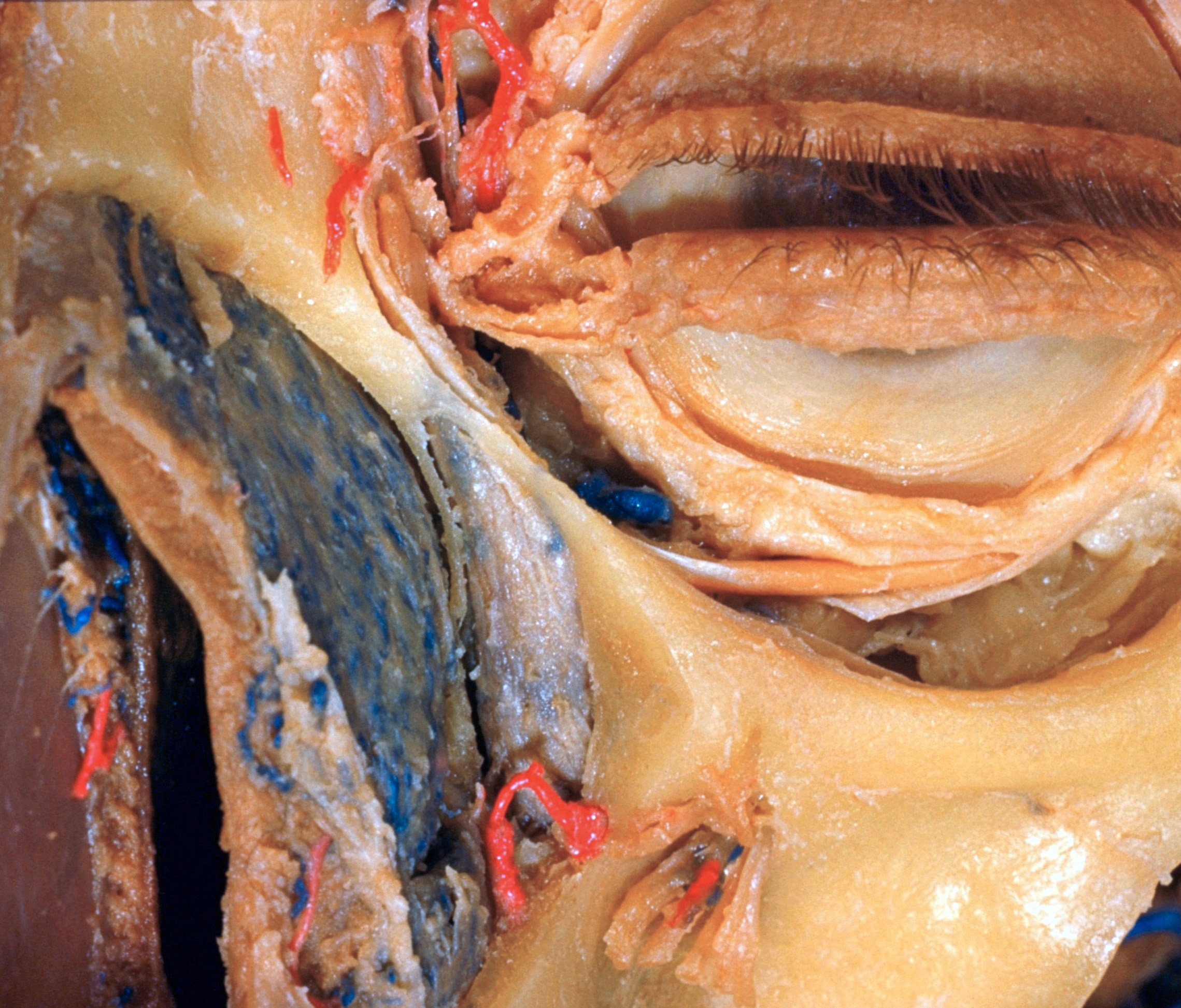
Dissection of left orbit from an anterior approach
Lacrimal sac; bony nasolacrimal canal opened
Stanford holds the copyright to the David L. Bassett anatomical images and has assigned
Creative Commons license Attribution-Share
Alike 4.0 International to all of the images.
For additional information regarding use and permissions,
please contact the Medical History Center.
Image #53-5
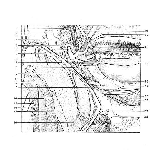
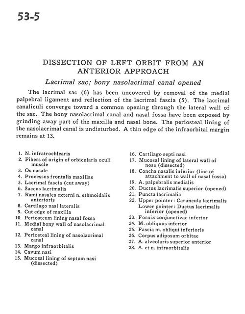
Dissection of left orbit from an anterior approach
Lacrimal sac; bony nasolacrimal canal opened
The lacrimal sac (6) has been uncovered by removal of the medial palpebral ligament and reflection of the lacrimal fascia (5). The lacrimal canaliculi converge toward a common opening through the lateral wall of the sac. The bony nasolacrimal canal and nasal fossa have been exposed by grinding away part of the maxilla and nasal bone. The periosteal lining of the nasolacrimal canal is undisturbed. A thin edge of the infraorbital margin remains at 13.
- Infratrochlear nerve
- Fibers of origin of orbicularis oculi muscle
- Nasal bone
- Frontal process of maxilla
- Lacrimal fascia (cut away)
- Lacrimal sac
- External nasal branches anterior ethmoidal nerve
- Lateral nasal cartilage
- Cut edge of maxilla
- Periosteum lining nasal fossa
- Medial bony wall of nasolacrimal canal
- Periosteal lining of nasolacrimal canal
- Infraorbital margin
- Nasal cavity
- Mucosal lining of nasal septum (dissected)
- Septal cartilage
- Mucosal lining of lateral wall of nose (dissected)
- Inferior nasal concha (line of attachment to wall of nasal fossa)
- Middle palpebral artery
- Superior lacrimal duct (opened)
- Lacrimal puncta
- Upper pointer: Lacrimal caruncle Lower pointer: Inferior lacrimal duct (opened)
- Inferior conjunctival fornix
- Inferior oblique muscle
- Fascia of inferior oblique muscle
- Orbital fat pad
- Anterior superior alveolar artery
- Infraorbital artery and nerve