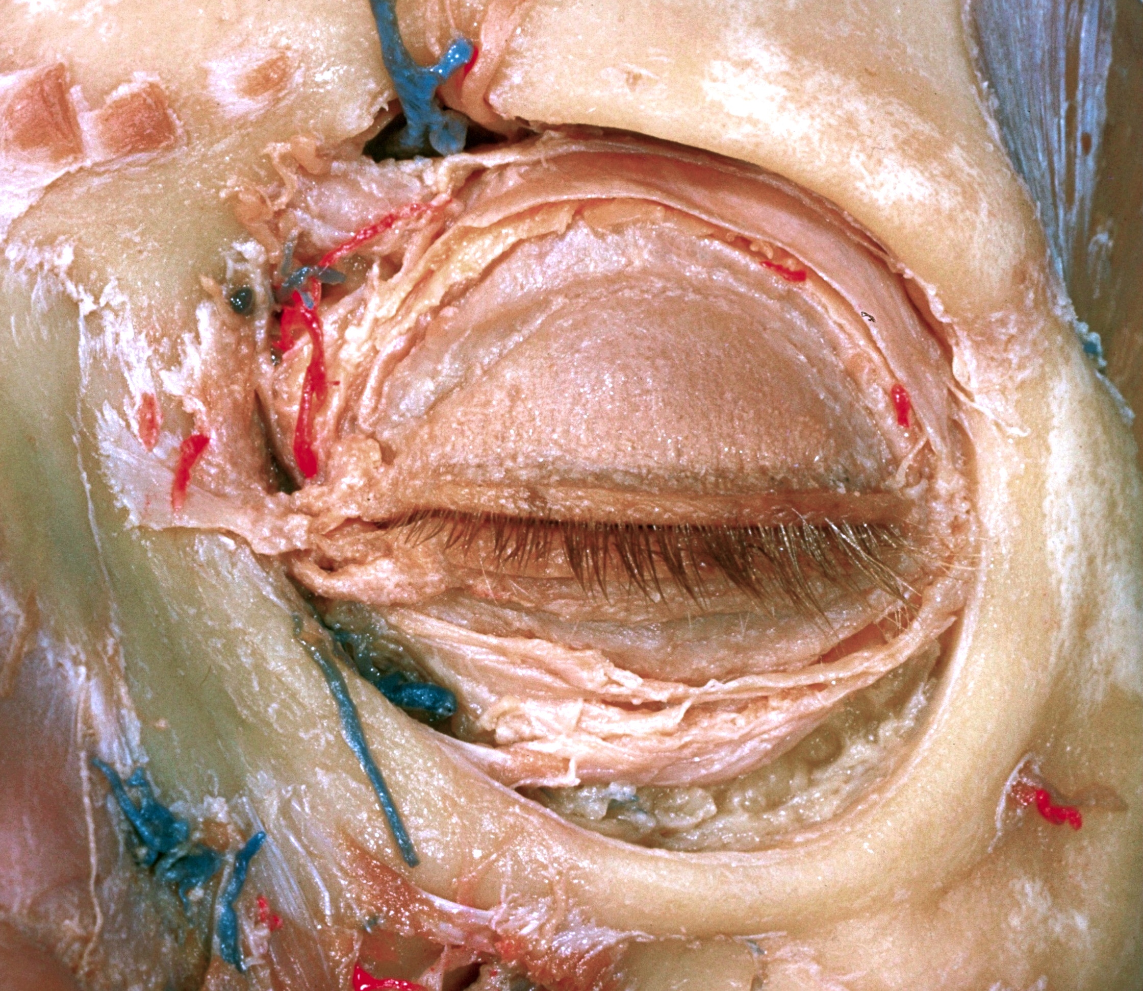
Dissection of left orbit from an anterior approach
Tarsal plates; lacrimal canaliculi; fascia supporting eye
Stanford holds the copyright to the David L. Bassett anatomical images and has assigned
Creative Commons license Attribution-Share
Alike 4.0 International to all of the images.
For additional information regarding use and permissions,
please contact the Medical History Center.
Image #53-3
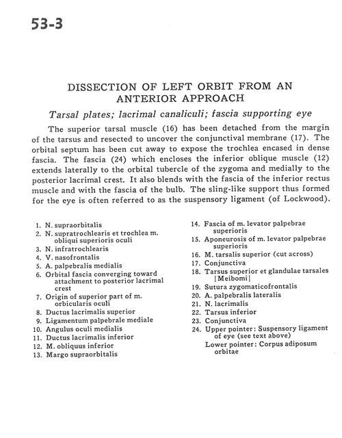
Dissection of left orbit from an anterior approach
Tarsal plates; lacrimal canaliculi; fascia supporting eye
The supporting tarsal muscle (16) has been detached from the margin of the tarsus and resected to uncover the conjunctival membrane (17). The orbital septum has been cut away to expose the trochlea encased in dense fascia. The fascia (24) which encloses the inferior oblique muscle (12) extends laterally to the orbital tubercle of the zygoma and medially to the posterior lacrimal crest. It also blends with the fascia of the inferior rectus muscle and with the fascia of the bulb. The sling-like support thus formed for the eye is often referred to as the suspensory ligament (of Lockwood).
- Supraorbital nerve
- Supratrochlear nerve and superior oblique muscle
- Infratrochlear nerve
- Nasofrontal vein
- Middle palpebral artery
- Orbital fascia converging toward attachment to posterior lacrimal crest
- Origin of superior part of orbicularis oculi muscle
- Superior lacrimal duct
- Medial palpebral ligament
- Medial ocular angle
- Inferior lacrimal duct
- Inferior oblique muscle
- Supraorbital margin
- Fascia of levator palpebrae superioris muscle
- Aponeurosis of levator palpebrae superioris muscle
- Superior tarsalis muscle (cut across)
- Conjunctiva
- Superior tarsus and tarsal (Meibomian) glands
- Zygomaticofrontal suture
- Lateral palpebral artery
- Lacrimal nerve
- Inferior tarsus
- Conjunctiva
- Upper pointer: Suspensory ligament of eye (see text above) Lower pointer: fat pad of orbit