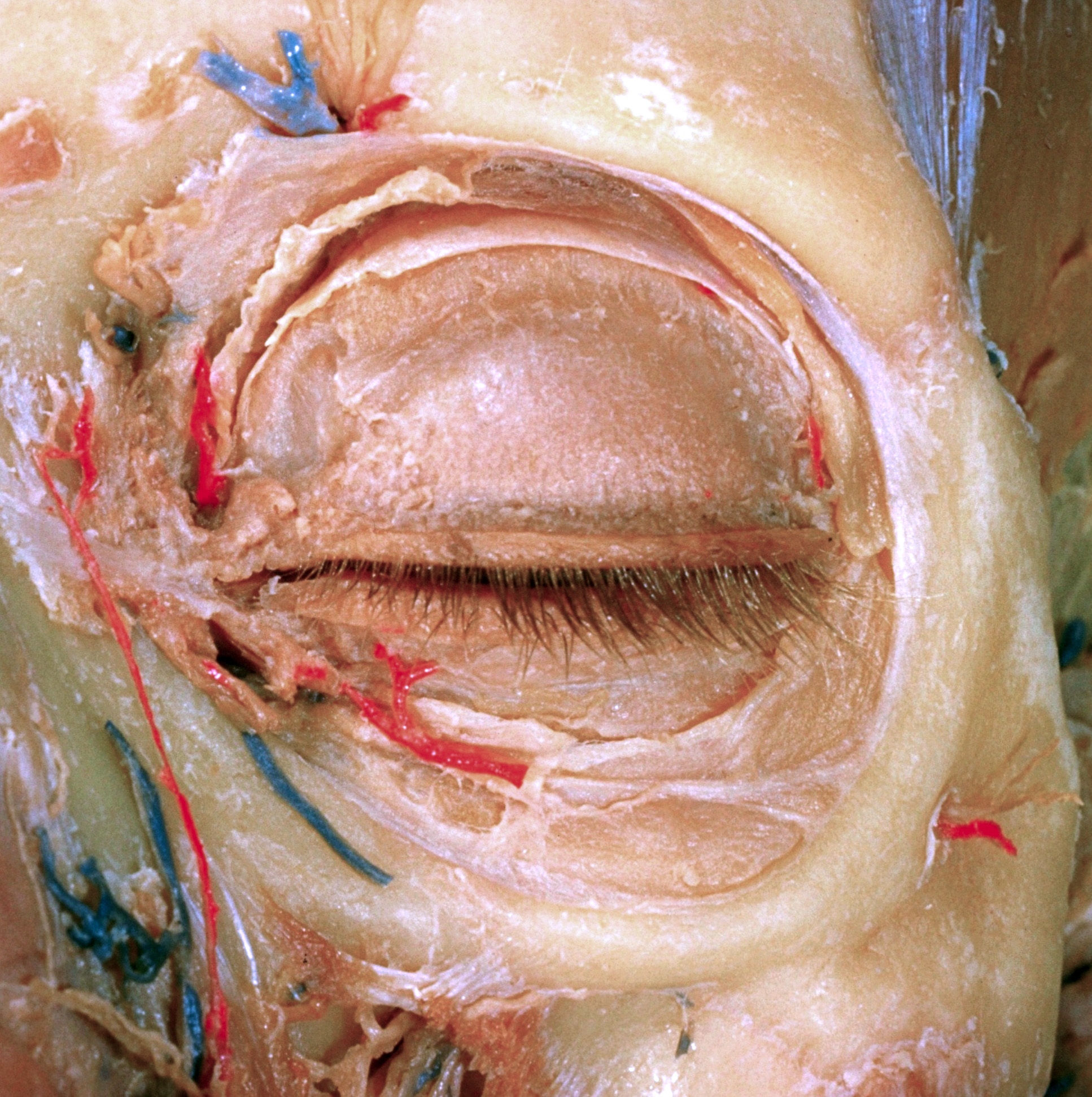
Dissection of left orbit from an anterior approach
Relation of orbital septum to aponeurotic and smooth muscle insertions of levator palpebrae superioris muscle
Stanford holds the copyright to the David L. Bassett anatomical images and has assigned
Creative Commons license Attribution-Share
Alike 4.0 International to all of the images.
For additional information regarding use and permissions,
please contact the Medical History Center.
Image #53-1

Dissection of left orbit from an anterior approach
Relation of orbital septum to aponeurotic and smooth muscle insertions of levator palpebrae superioris muscle
The aponeurosis of the levator palpebrae superioris muscle has been cut back so that the entire extent of the tarsal muscle is visible. Smooth muscle fibers are less evident in the extreme lateral and medial parts of this layer. The orbital septum does not appear to be a complete membrane in the lateral and inferior parts of the orbit, but rather consists of fibrous bands intermingled with lobules of fat. The layer of connective tissue which extended betweeen the orbicularis oculi muscle and the orbital septum has been completely removed.
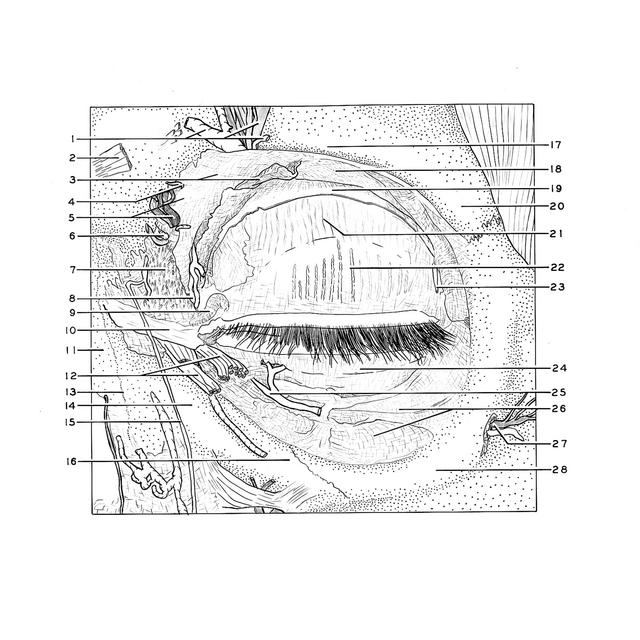
- Supraorbital nerve, artery, and vein
- Remnant of origin of corrugator supercilli muscle
- Orbital septum (detached from aponeurosis of levator palpebrae muscle)
- Supratrochlear nerve
- Upper pointer: Position of trochlea Lower pointer: Infratrochlear nerve
- Nasofrontal vein emerging from orbit
- Area of origin of upper part of orbicularis oculi muscle
- Middle palpebral artery
- Superior canaliculus
- Medial palpebral ligament
- Nasal bone
- Fibers of orbicularis oculi muscle (cut across near origin)
- Nasomaxillary suture
- Frontal process of maxilla
- Small artery along periosteum
- Zygomaticomaxillary suture
- Supraorbital margin of orbit
- Fascia of levator palpebrae superioris muscle
- Aponeurosis of Ievator palpebrae superioris muscle
- Zygomatic process frontal bone
- Superior tarsalis muscle (muscle of Müller)
- Superior tarsus and tarsal (Meibomian) glands
- Lateral palpebral artery
- Inferior tarsus
- Orbital branch (aberrant) from external maxillary artery
- Orbital septum
- Zygomatico-facial branch of zygomatic nerve
- Zygomatic bone