Dissection of left orbit from an anterior approach
Orbital septum; tarsal plates; insertion of levator palpebrae superioris muscle
Stanford holds the copyright to the David L. Bassett anatomical images and has assigned
Creative Commons license Attribution-Share
Alike 4.0 International to all of the images.
For additional information regarding use and permissions,
please contact the Medical History Center.
Image #52-6
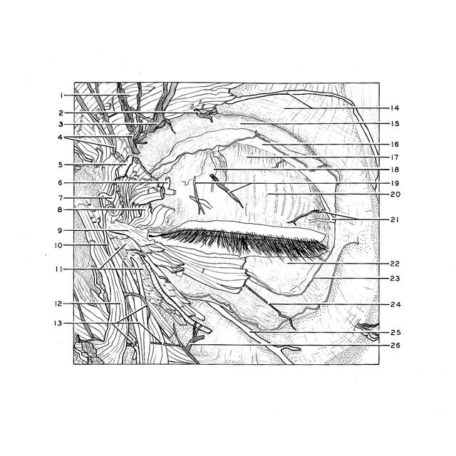
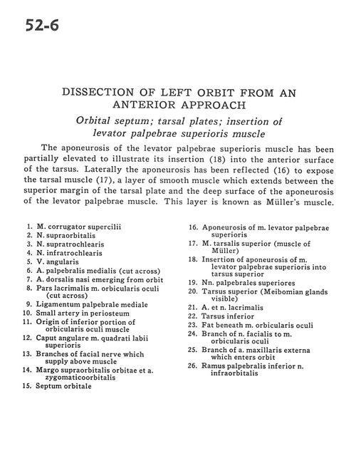
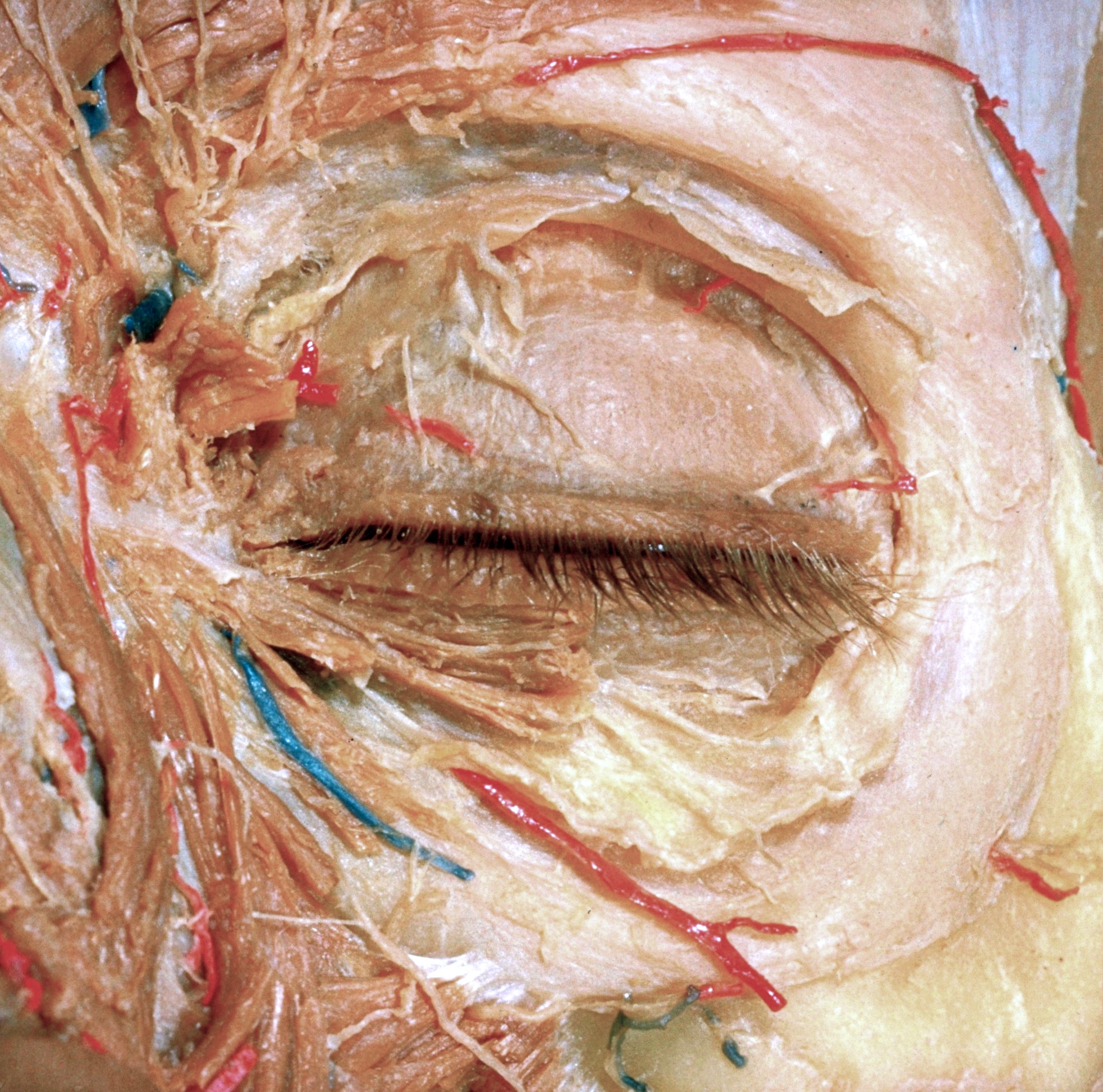
Dissection of left orbit from an anterior approach
Orbital septum; tarsal plates; insertion of levator palpebrae superioris muscle
The aponeurosis of the levator palpebrae superioris muscle has been partially elevated to illustrate its insertion (18) into the anterior surface of the tarsus. Laterally the aponeurosis has been reflected (16) to expose the tarsal muscle (17), a layer of smooth muscle which extends between the superior margin of the tarsal plate and the deep surface of the aponeurosis of the levator palpebrae muscle. This layer is known as M|ller's muscle.
- Corrugator supercilii muscle
- Supraorbital nerve
- Supratrochlear nerve
- Infratrochlear nerve
- Angular vein
- Middle palpebral artery (cut across)
- Dorsal nasal artery emerging from orbit
- Lacrimal part of orbicularis oculi muscle (cut across)
- Medial palpebral ligament
- Small artery in periosteum
- Origin of inferior portion of orbicularis oculi muscle
- Angular head of levator labii superioris muscle
- Branches of facial nerve which supply above muscle
- Supraorbital margin and zygomatico-orbital artery
- Orbital septum
- Aponeurosis of levator palpebrae superioris muscle
- Superior tarsal muscle (muscle of Müller)
- Insertion of aponeurosis of levator palpebrae superioris muscle into superior tarsus
- Superior palpebral nerve
- Superior tarsus (Meibomian glands visible)
- Lacrimal nerve and artery
- Inferior tarsus
- Fat beneath orbicularis oculi muscle
- Branch of facial nerve to orbicularis oculi muscle
- Branch of external maxillary artery which enters orbit
- Inferior palpebral branch of infraorbital nerve