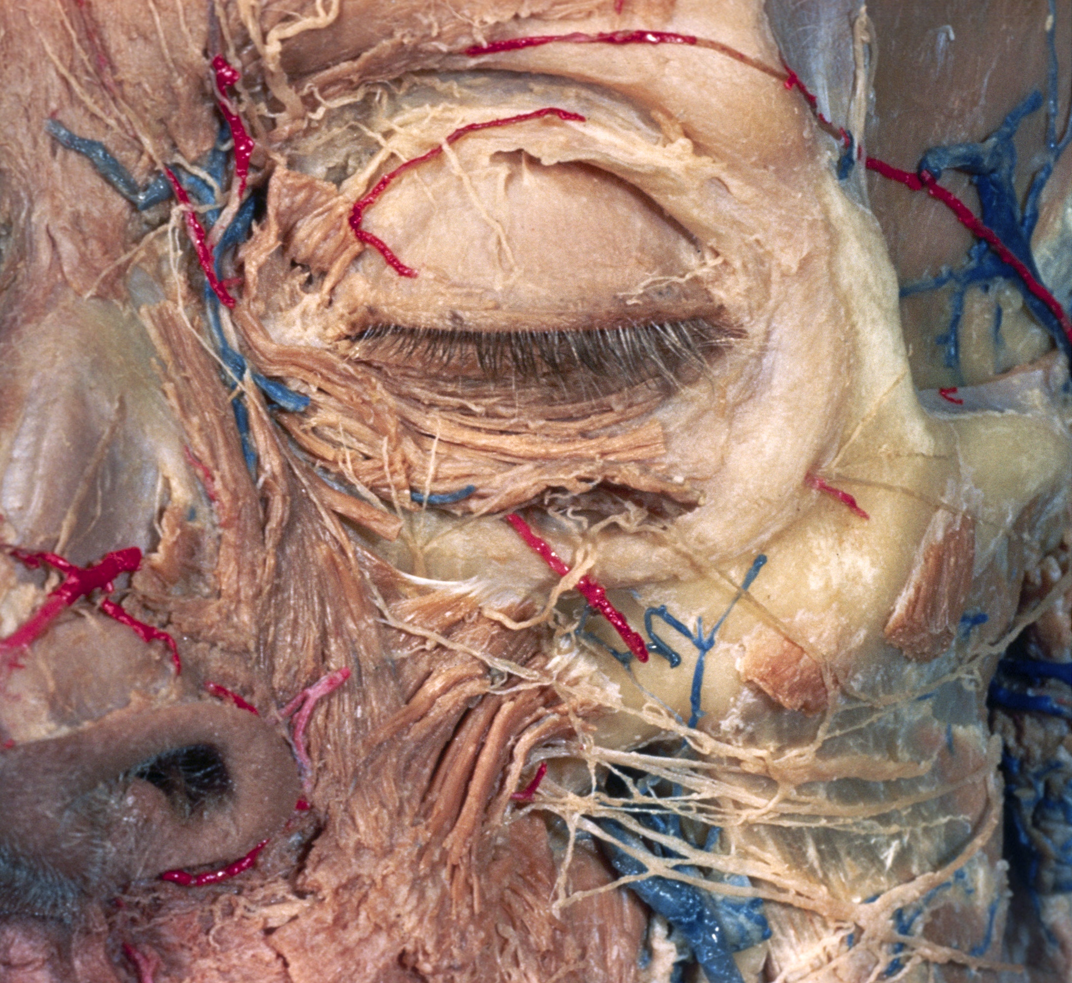
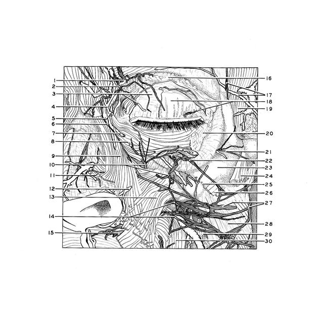
Dissection of left orbit from an anterior approach
Branches of trigeminal and facial nerves in orbital and infraorbital regions
Stanford holds the copyright to the David L. Bassett anatomical images and has assigned
Creative Commons license Attribution-Share
Alike 4.0 International to all of the images.
For additional information regarding use and permissions,
please contact the Medical History Center.
Image #52-5
Dissection of left orbit from an anterior approach
Branches of trigeminal and facial nerves in orbital and infraorbital regions
Portions of the orbicularis oculi muscle remain over the medial parts of the eyelids but have been reflected in various ways to show underlying structures. The zygomatic muscle (24) and zygomatic head of the quadratus labii superioris muscle (26) have been partially removed.
- Supratrochlear nerve
- Infratrochlear nerve
- Upper pointer: Fascia over superior tarsus Lower pointer: Medial palpebral artery (branch which communicates with supraorbital artery displaced inferiorly)
- Ascending branches of dorsal nasal artery
- Dorsal nasal artery emerging from orbit
- Anastomotic branch between infratrochlear nerve and facial nerve
- Angular vein (divided)
- Orbicularis oculi muscle (reflected upward)
- Angular head of levator labii superioris muscle
- Inferior palpebral branches of infraorbital nerve
- External nasal branch infraorbital nerve
- Alar branch of angular artery
- Infraorbital head of levator labii superioris muscle
- Communications between buccal branches of facial nerve and superior labial branches of infraorbital nerve
- Superior labial branches of infraorbital nerve
- Orbital septum
- Zygomaticotemporal branch zygomatic nerve and zygomatico-orbital artery
- Superior tarsus and lacrimal nerve
- Temporalis muscle
- Layer of fat beneath orbicularis oculi muscle
- Zygomaticofacial branch of zygomatic nerve
- Communication between zygomatic branch facial nerve and inferior palpebral branch of infraorbital nerve
- Malar surface zygomatic bone
- Zygomaticus muscle (cut across)
- Zygomatico-maxillary suture
- Zygomatic head of levator labii superioris muscle
- Buccal branches of facial nerve
- Masseter muscle
- Parotid duct (cut across)
- Buccinator muscle