Dissection of left orbit from an anterior approach
Insertion of levator palpebrae superioris muscle
Stanford holds the copyright to the David L. Bassett anatomical images and has assigned
Creative Commons license Attribution-Share
Alike 4.0 International to all of the images.
For additional information regarding use and permissions,
please contact the Medical History Center.
Image #52-3
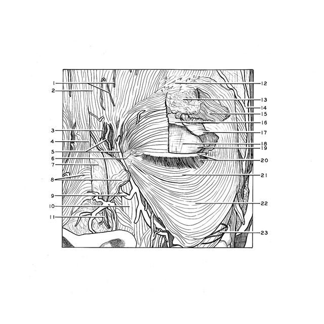
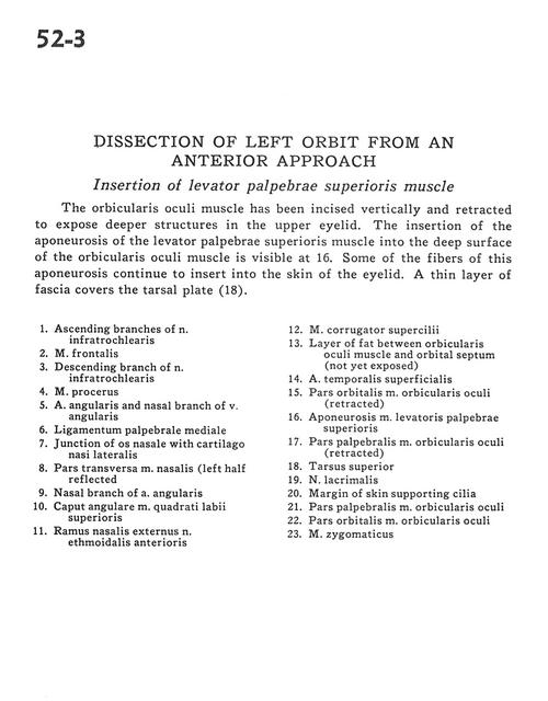
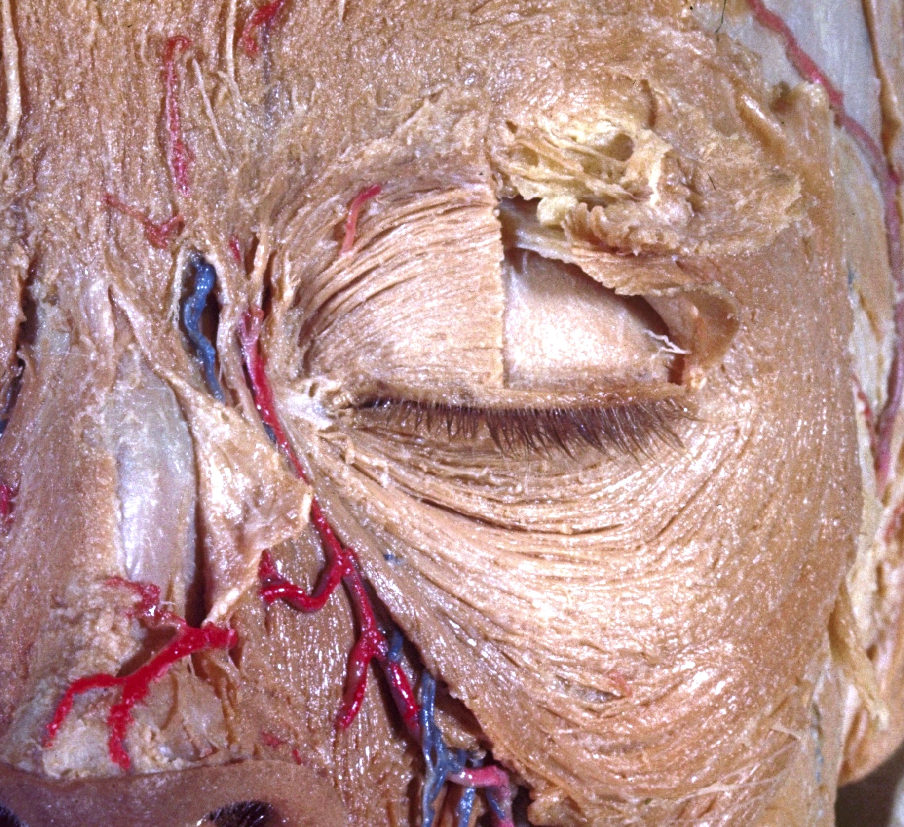
Dissection of left orbit from an anterior approach
Insertion of levator palpebrae superioris muscle
The orbicularis oculi muscle has been incised vertically and retracted to expose deeper structures in the upper eyelid. The insertion of the aponeurosis of the levator palpebrae superioris muscle into the deep surface of the orbicularis oculi muscle is visible at 16. Some of the fibers of this aponeurosis continue to insert into the skin of the eyelid. A thin layer of fascia covers the tarsal plate (18).
- Ascending branches of infratrochlear nerve
- Frontalis muscle
- Descending branch of infratrochlear nerve
- Procerus muscle
- Angular artery and nasal branch of angular vein
- Middle palpebral ligament
- Junction of nasal bone with lateral nasal cartilage
- Transverse part of nasalis muscle (left half reflected)
- Nasal branch of angular artery
- Angular head of levator labii superioris muscle
- External nasal branch of anterior ethmoidal nerve
- Corrugator supercilii muscle
- Layer of fat between orbicularis oculi muscle and orbital septum (not yet exposed)
- Superficial temporal artery
- Orbital part orbicularis oculi muscle (retracted)
- Aponeurosis levator palpebrae superioris muscle
- Palpebral part of orbicularis oculi muscle (retracted)
- Superior tarsus
- Lacrimal nerve
- Margin of skin supporting cilia
- Palpebral part of orbicularis oculi muscle
- Orbital part orbicularis oculi muscle
- Zygomaticus muscle