Floor of cranial cavity
Structures inferior to left middle cranial fossa, superior view
Stanford holds the copyright to the David L. Bassett anatomical images and has assigned
Creative Commons license Attribution-Share
Alike 4.0 International to all of the images.
For additional information regarding use and permissions,
please contact the Medical History Center.
Image #51-4

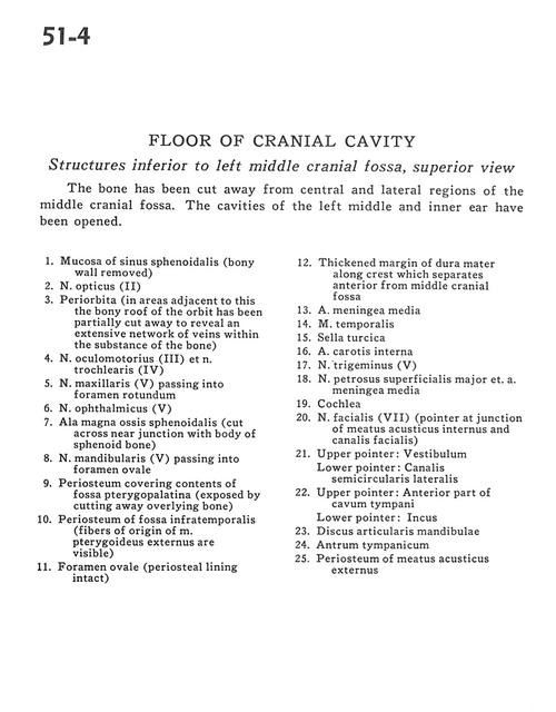
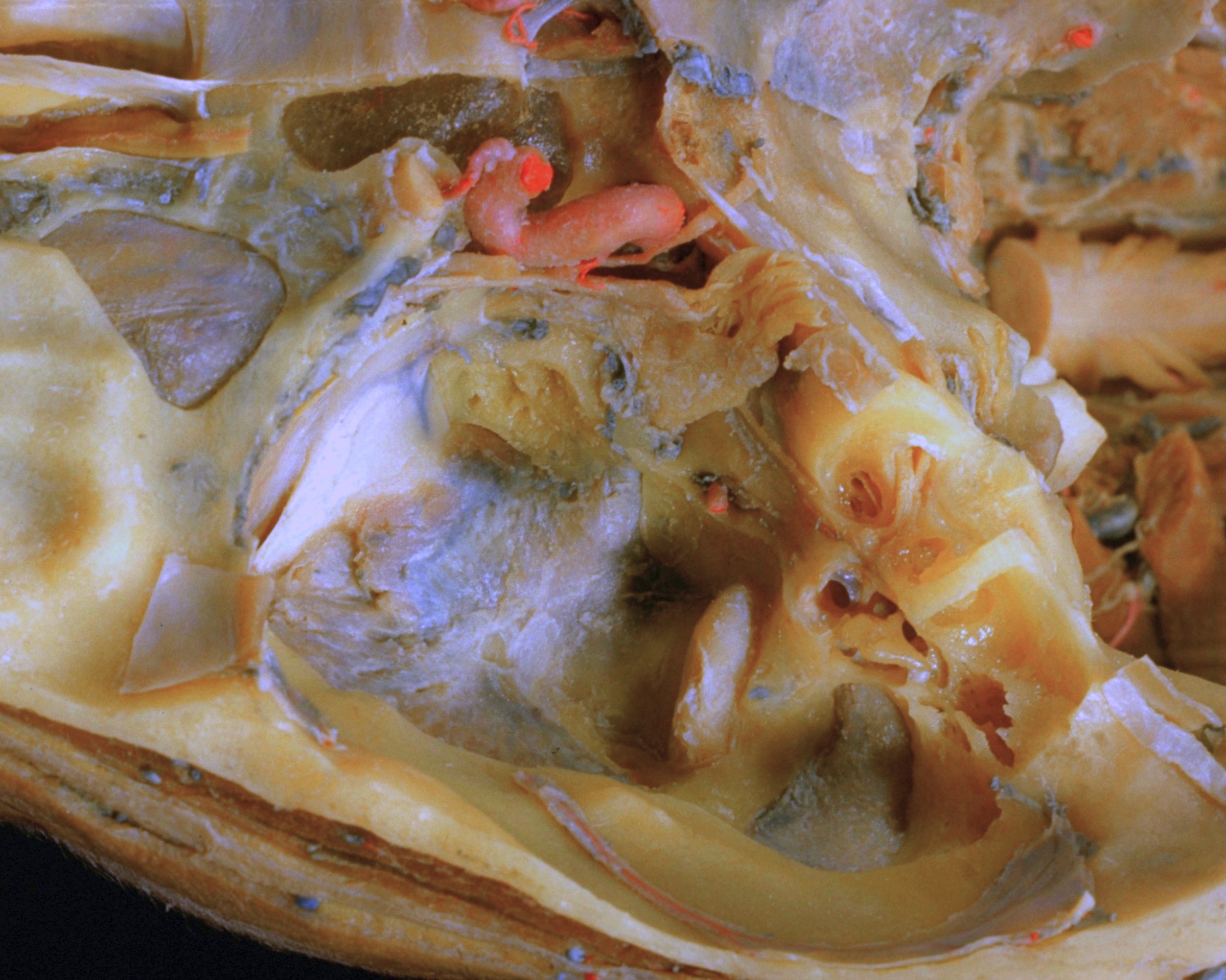
Floor of cranial cavity
Structures inferior to left middle cranial fossa, superior view
The bone has been cut away from central and lateral regions of the middle cranial fossa. The cavities of the left middle and inner ear have been opened.
- Mucosa of sphenoid sinus (bony wall removed)
- Optic nerve (II)
- Periorbita (in areas adjacent to this the bony roof of the orbit has been partially cut away to reveal an extensive network of veins within the substance of the bone)
- Oculomotor nerve (III) and trochlear nerve (IV)
- Maxillary nerve (V2) passing into foramen rotundum
- Ophthalmic nerve (Vl)
- Greater wing of sphenoid (cut across near junction with body of sphenoid bone)
- Mandibular nerve (V3) passing into foramen ovale
- Periosteum covering contents of pterygopalatine fossa (exposed by cutting away overlying bone)
- Periosteum of infratemporal fossa (fibers of origin of external pterygoid muscle are visible)
- Foramen ovale (periosteal lining intact)
- Thickened margin of dura mater along crest which separates anterior from middle cranial fossa
- Middle meningeal artery
- Temporalis muscle
- Sella turcica
- Internal carotid artery
- Trigeminal nerve (V)
- Major superficial petrous nerve and middle meningeal artery
- Cochlea
- Facial nerve (VII) (pointer at junction of internal acoustic meatus and facial canal)
- Upper pointer: Vestibule Lower pointer: Semicircular canal (lateral)
- Upper pointer: Anterior part of tympanic cavity Lower pointer: Incus
- Articular disc of mandible
- Tympanic antrum
- Periosteum of external acoustic meatus