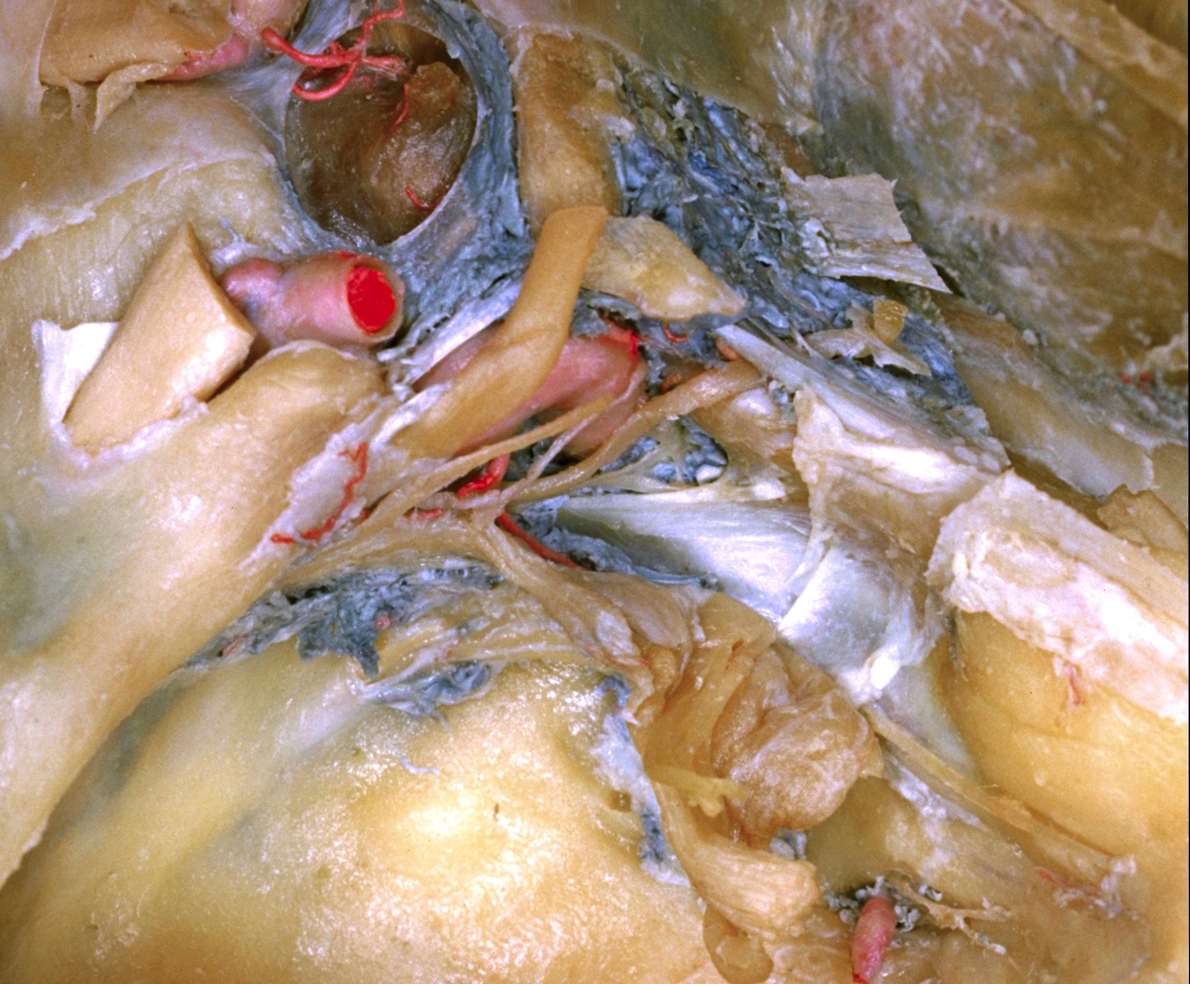
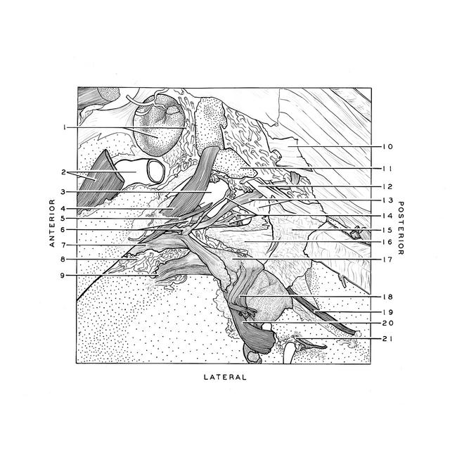
Floor of cranial cavity
Middle cranial fossa; dissection of left cavernous sinus (continued); portio minor of trigeminal nerve; abducens nerve; carotid plexus
Stanford holds the copyright to the David L. Bassett anatomical images and has assigned
Creative Commons license Attribution-Share
Alike 4.0 International to all of the images.
For additional information regarding use and permissions,
please contact the Medical History Center.
Image #51-2
Floor of cranial cavity
Middle cranial fossa; dissection of left cavernous sinus (continued); portio minor of trigeminal nerve; abducens nerve; carotid plexus
The trigeminal nerve has been turned laterally to expose its motor root (18), the fibrous tissue partially closing the foramen lacerum (16), and the relations of the carotid artery and cavernous plexus of nerves (14).
- Circular sinus and hypophysis
- Internal carotid artery and optic nerve (II)
- Internal carotid artery within cavernous sinus
- Oculomotor nerve (III)
- Trochlear nerve (IV)
- Upper pointer: Branch of internal carotid artery to meninges and semilunar ganglion Lower pointer: Communication between internal carotid sympathetic plexus and ophthalmic nerve
- Ophthalmic nerve (V1)
- Cavernous sinus near junction with superior ophthalmic vein
- Maxillary nerve (V2)
- Posterior longitudinal ligament
- Posterior clinoid process
- Petroclinoid ligament
- Upper pointer: Abducens nerve (VI) Lower pointer: Apex of pyramid
- Cavernous nerve plexus
- Dura mater forming medial wall of trigeminal (Meckel's) cave
- Dura mater bridging across sphenopetrous fissure
- Semilunar ganglion
- Minor portion trigeminal nerve (V)
- Greater superficial petrosal nerve
- Major portion of trigeminal nerve (V) (reflected laterally)
- Lesser superficial petrosal nerve