Floor of cranial cavity
Structures in region of sella turcica; origin of ophthalmic arteries, superior view
Stanford holds the copyright to the David L. Bassett anatomical images and has assigned
Creative Commons license Attribution-Share
Alike 4.0 International to all of the images.
For additional information regarding use and permissions,
please contact the Medical History Center.
Image #50-6
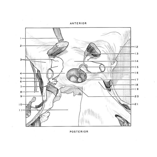
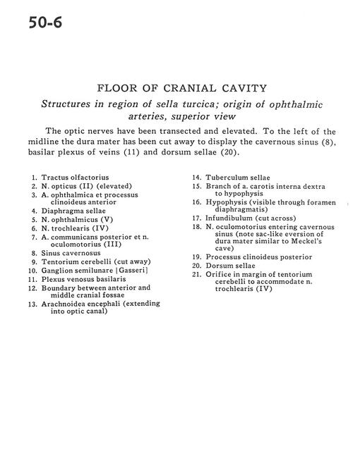
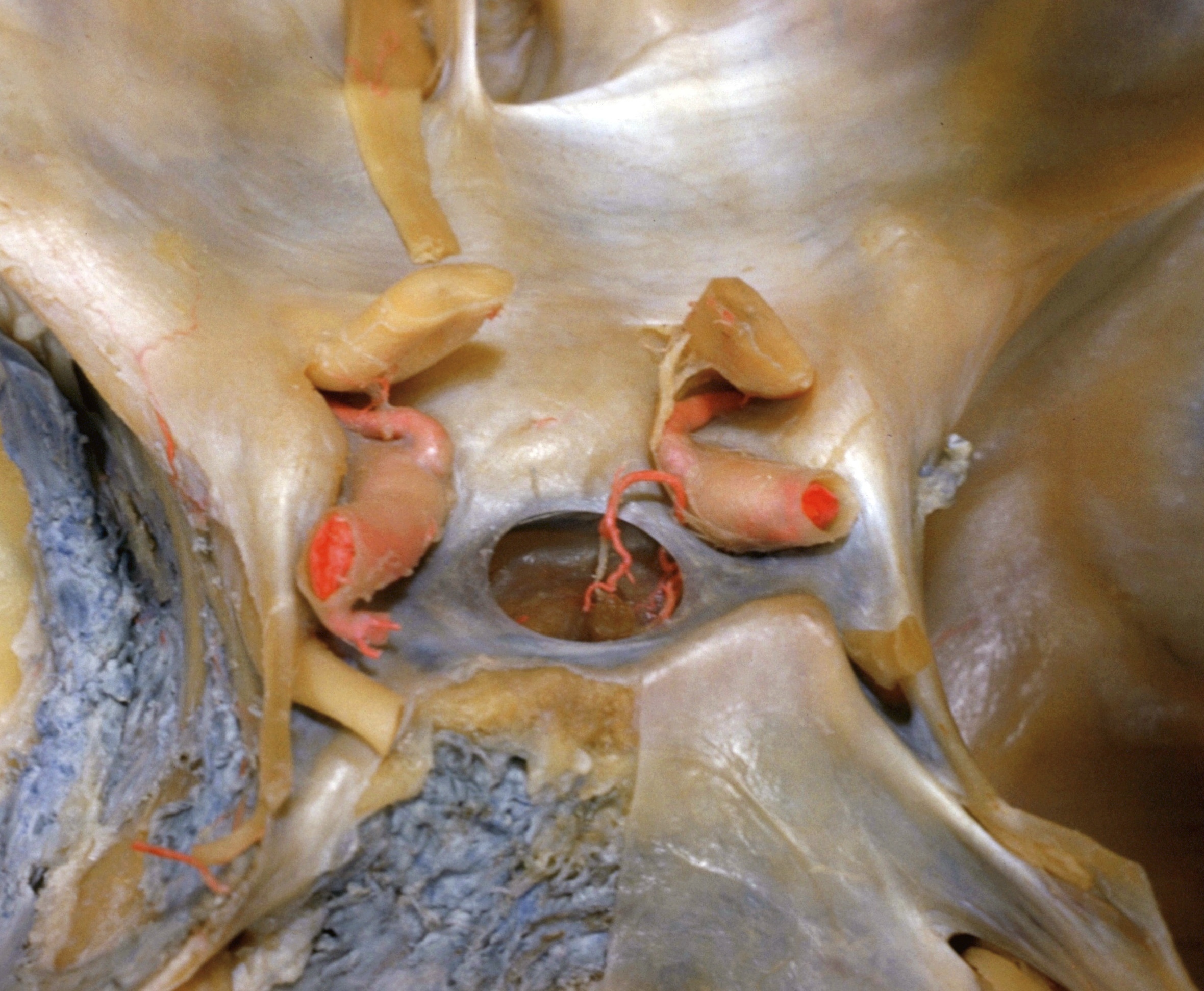
Floor of cranial cavity
Structures in region of sella turcica; origin of ophthalmic arteries, superior view
The optic nerves have been transected and elevated. To the left of the midline the dura mater has been cut away to display the cavernous sinus (8), basilar plexus of veins (11) and dorsum sellae (20).
- Olfactory tract
- Optic nerve (II) (elevated)
- Ophthalmic artery and anterior clinoid process
- Diaphragm of sella
- Ophthalmic nerve (VI)
- Trochlear nerve (IV)
- Posterior communicating artery and oculomotor nerve (III)
- Cavernous sinus
- Tentorium cerebelli (cut away)
- Semilunar ganglion (trigeminal)
- Basilar venous plexus
- Boundary between anterior and middle cranial fossae
- Arachnoid matter (extending into optic canal)
- Tuberculum sellae
- Branch of internal carotid artery right to hypophysis
- Hypophysis (visible through diaphragmatic foramen)
- Infundibulum (cut across)
- Oculomotor nerve entering cavernous sinus (note sac-like eversion of dura mater similar to Meckel's cave)
- Posterior clinoid process
- Dorsum sellae
- Orifice in margin of tentorium cerebelli to accommodate trochlear nerve (IV)