General orientation views of dissection
Deep facial dissection, left lateral view; internal maxillary artery
Stanford holds the copyright to the David L. Bassett anatomical images and has assigned
Creative Commons license Attribution-Share
Alike 4.0 International to all of the images.
For additional information regarding use and permissions,
please contact the Medical History Center.
Image #49-6
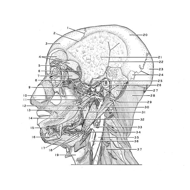
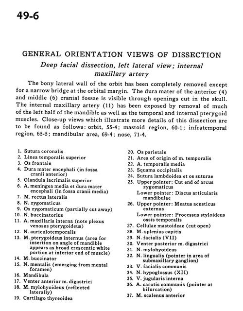
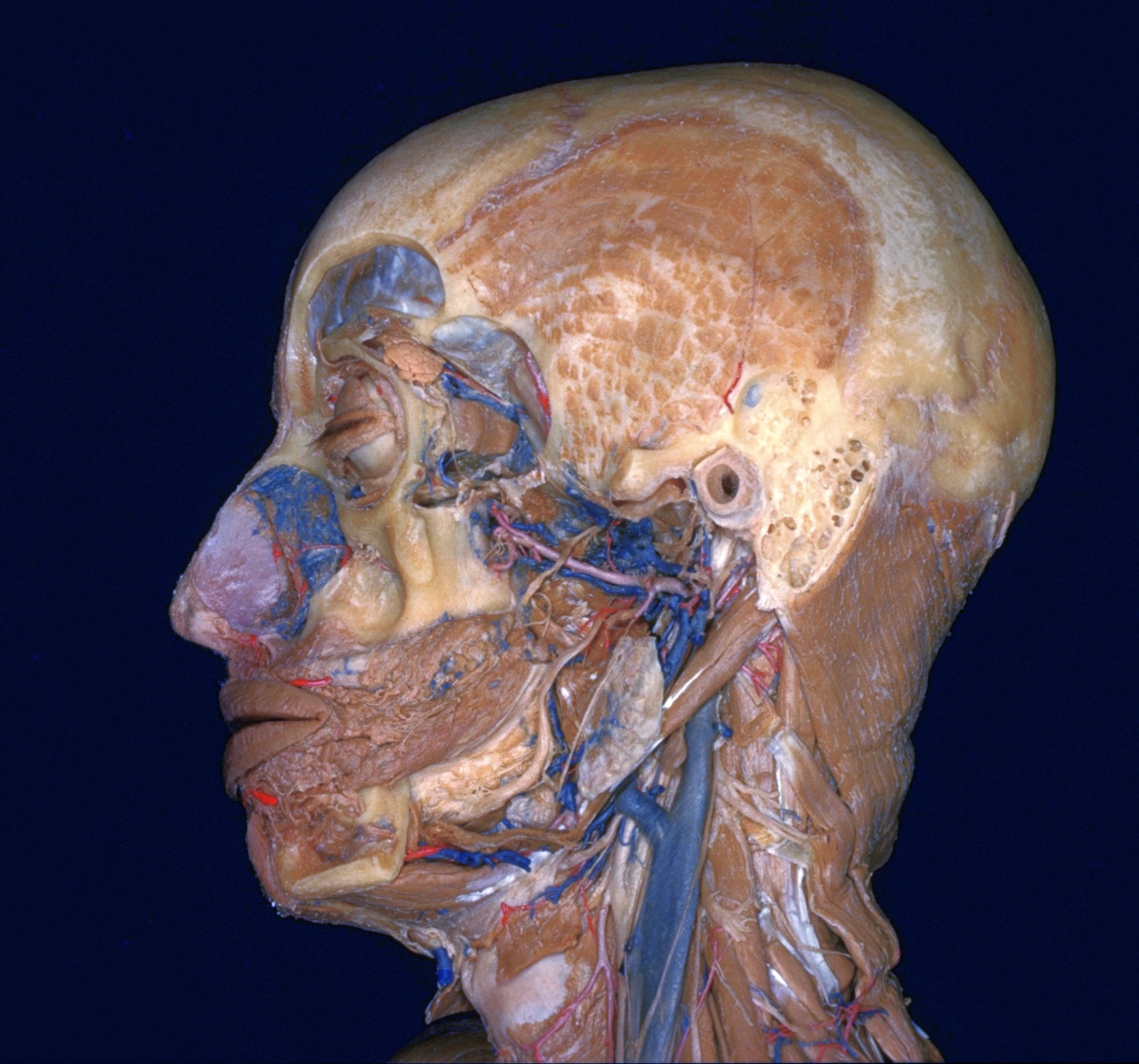
General orientation views of dissection
Deep facial dissection, left lateral view; internal maxillary artery
The bony lateral wall of the orbit has been completely removed except for a narrow bridge at the orbital margin. The dura mater of the anterior (4) the middle (6) cranial fossae is visible through openings cut in the skull. The internal maxillary artery (11) has been exposed by removal of much of the left half of the mandible as well as the temporal and internal pterygoid muscles. Close-up views which illustrate more details of this dissection are to be found as follows
- Coronal suture
- Superior temporal line
- Frontal bone
- Dura mater encephali (in anterior cranial fossa)
- Superior lacrimal gland
- Middle meningeal artery and dura mater encephali (in middle cranial fossa)
- Lateral rectus muscle
- Zygomatic nerve
- Zygomatic bone (partially cut away)
- Buccal nerve
- Internal maxillary artery (note plexus venosus pterygoideus)
- Auriculotemporal nerve
- Internal pterygoid muscle (area for insertion on angle of mandible appears as broad crescentic white portion at inferior end of muscle)
- Buccinator muscle
- Mental nerve (emerging from mental foramen)
- Mandible
- Anterior belly digastric muscle
- Mylohyoid muscle (reflected laterally)
- Thyroid cartilage
- Parietal bone
- Area of origin of temporalis muscle
- Middle temporal artery
- Occipital bone (squamous part)
- Lambdoidal suture and intersutural bone
- Upper pointer: Cut end of zygomatic arch Lower pointer: Articular disc for mandible
- Upper pointer: External acoustic meatus Lower pointer: Styloid process temporal bone
- Mastoid cells (cut open)
- Splenius capitis muscle
- Facial nerve (VII)
- Posterior belly of digastric muscle
- Mylohyoid nerve
- Lingual nerve (pointer in area of submandibular ganglion)
- Common facial vein
- Hypoglossal nerve (Xll)
- Internal jugular vein
- Common carotid artery (pointer at bifurcation)
- Anterior scalene muscle