General orientation views of dissection
Deep facial dissections; masseter muscle reflected, anterolateral view
Stanford holds the copyright to the David L. Bassett anatomical images and has assigned
Creative Commons license Attribution-Share
Alike 4.0 International to all of the images.
For additional information regarding use and permissions,
please contact the Medical History Center.
Image #49-4
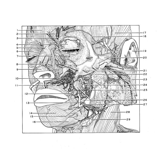
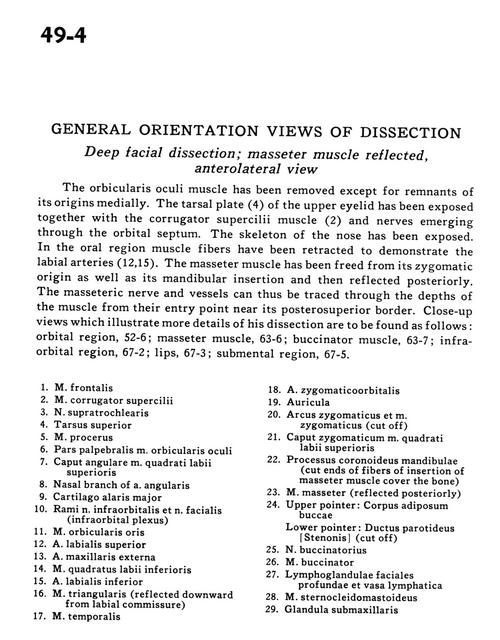
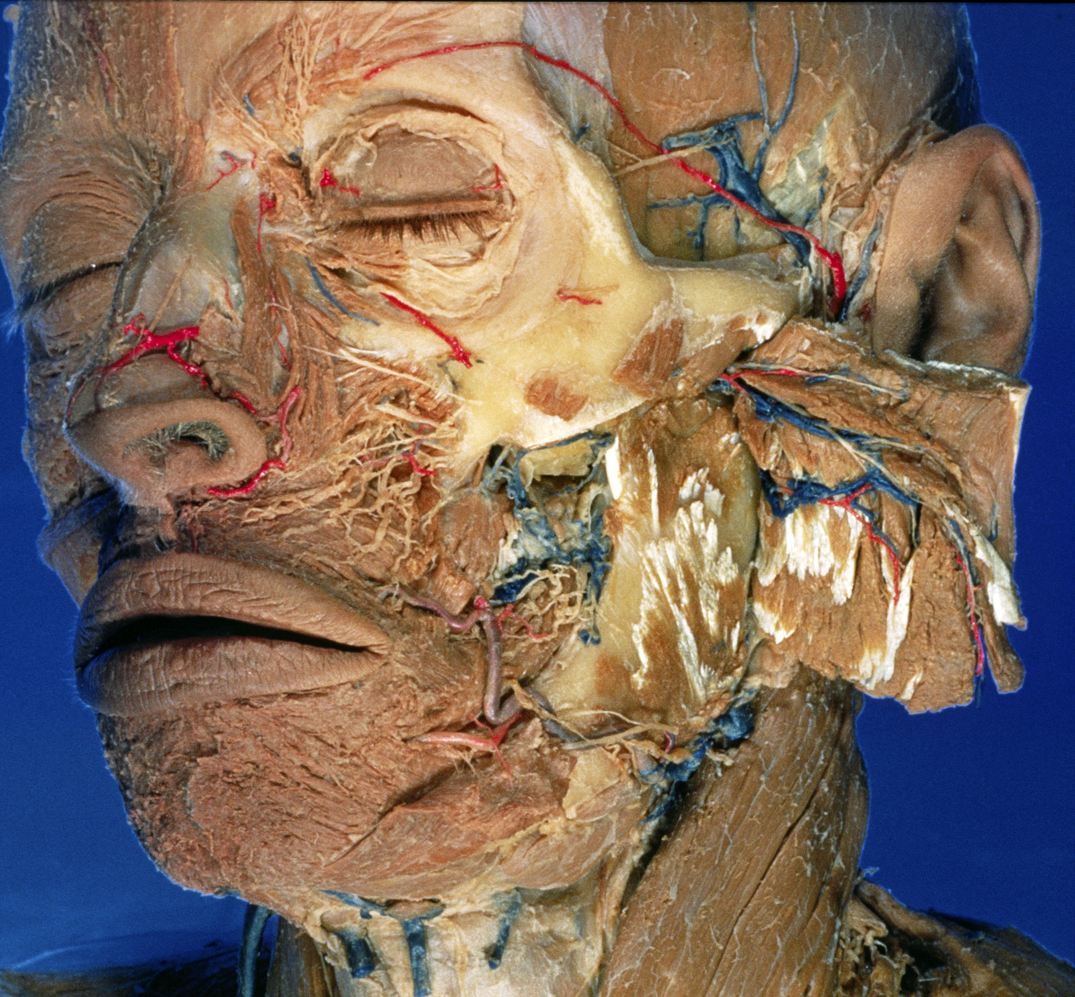
General orientation views of dissection
Deep facial dissections; masseter muscle reflected, anterolateral view
The orbicularis oculi muscle has been removed except for remnants of its origins medially. The tarsal plate (4) of the upper eyelid has been exposed together with the corrugator supercilii muscle (2) and nerves emerging through the orbital septum. The skeleton of the nose has been exposed. In the oral region muscle fibers have been retracted to demonstrate the labial arteries (12,15). The masseter muscle has been freed from its zygomatic origin as well as its mandibular insertion and then reflected posteriorly. The masseteric nerve and vessels can thus be traced through the depths of the muscle from their entry point near its posterosuperior border. Close-up views which illustrate more details of this dissection are to be found as follows
- Muscle frontalis
- Muscle corrugator supercilii
- Supratrochlear nerve
- Superior tarsus
- Muscle procerus
- Palpebral part of orbicularis oculi muscle
- Angular head of levator labii superioris muscle
- Nasal branch of angular artery
- Greater alar cartilage
- Branches of infraorbital nerve and facial nerve (infraorbital plexus)
- Muscle orbicularis otis
- Superior labial artery
- External maxillary artery
- Depressor labii inferioris muscle
- Inferior labial artery
- Depressor anguli oris (reflected downward from labial commissure)
- Muscle temporalis
- Zygomatico-orbital artery
- Auricle
- Zygomatic arch and zygomatic muscle (cut off)
- Head of zygomatic levator labii superioris muscle
- Coronoid process of mandible (cut ends of fibers of insertion of masseter muscle cover the bone)
- Muscle masseter (reflected posteriorly)
- Upper pointer: Body adiposum buccae Lower pointer: Parotid duct (cut off)
- Buccal nerve
- Muscle buccinator
- Deep facial lymph nodes and lymph vessels
- Sternocleidomastoid muscle
- Submandibular gland