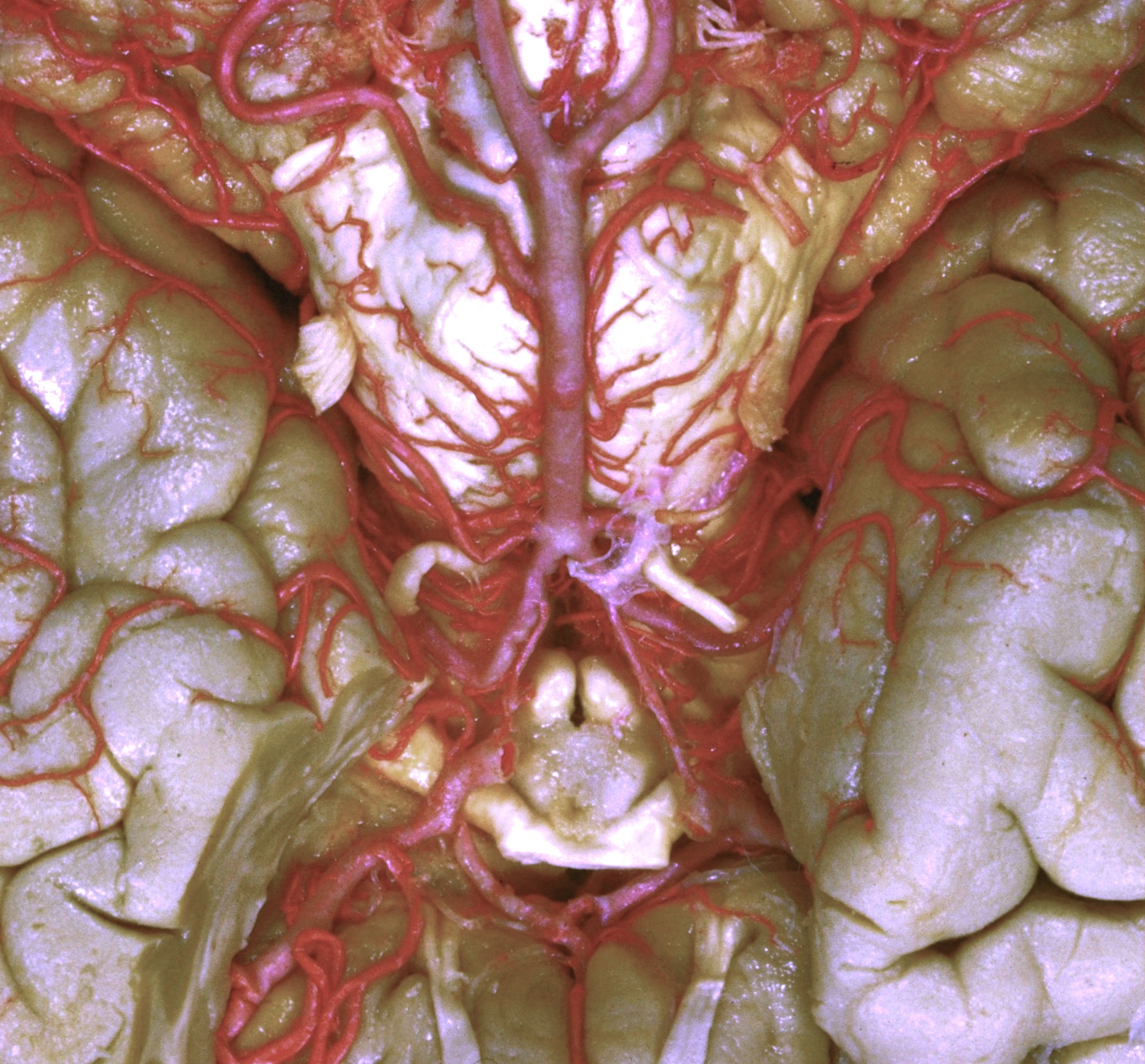
Exploration of the brain from its basal aspect
Arteries of basal surface of brain
Stanford holds the copyright to the David L. Bassett anatomical images and has assigned
Creative Commons license Attribution-Share
Alike 4.0 International to all of the images.
For additional information regarding use and permissions,
please contact the Medical History Center.
Image #4-4
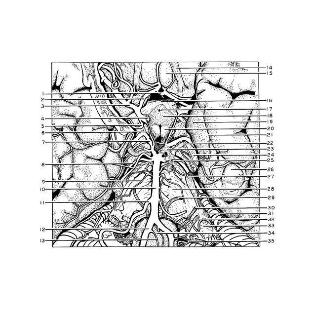
Exploration of the brain from its basal aspect
Arteries of basal surface of brain
The vertebral and internal carotid arteries are visible as they reach the basal surface of the brain to enter into the formation of the arterial circle of Willis. A portion of the right temporal lobe has been cut away to reveal the middle cerebral artery with some of its branches deep in the lateral fissure. The optic chiasm is partially removed to display the anterior communicating artery. The internal auditory arteries are not clearly demonstrated in this specimen.
- Anterior communicating artery
- Anterior cerebral artery
- Middle cerebral artery
- Internal carotid artery (cut across)
- Choroidal artery (anterior)
- Posterior communicating artery
- Posterior cerebral artery
- Superior cerebellar artery (double on right, single vessel on left)
- Branch to pons from basilar artery
- Basilar artery
- Anterior inferior cerebellar artery
- Vertebral artery
- Anterior spinal artery
- Olfactory tract left
- Straight gyrus
- Optic chiasm and optic tract
- Infundibulum and infundibular recess third ventricle
- Tuber cinereum
- Uncus
- Mamillary body
- Fissura collateralis
- Oculomotor nerve (III)
- Arachnoid (in situ)
- Cerebral peduncle
- Trochlear nerve (IV)
- Fusiform gyrus
- Trigeminal nerve (V)
- Pons
- Abducens nerve (VI)
- Facial nerve (VII)
- Middle cerebellar peduncle
- Vestibulocochlear nerve (VIII)
- Flocculus
- Medulla oblongata
- Glossopharyngeal nerves (IX) and vagus (X); Choroid plexus fourth ventricle