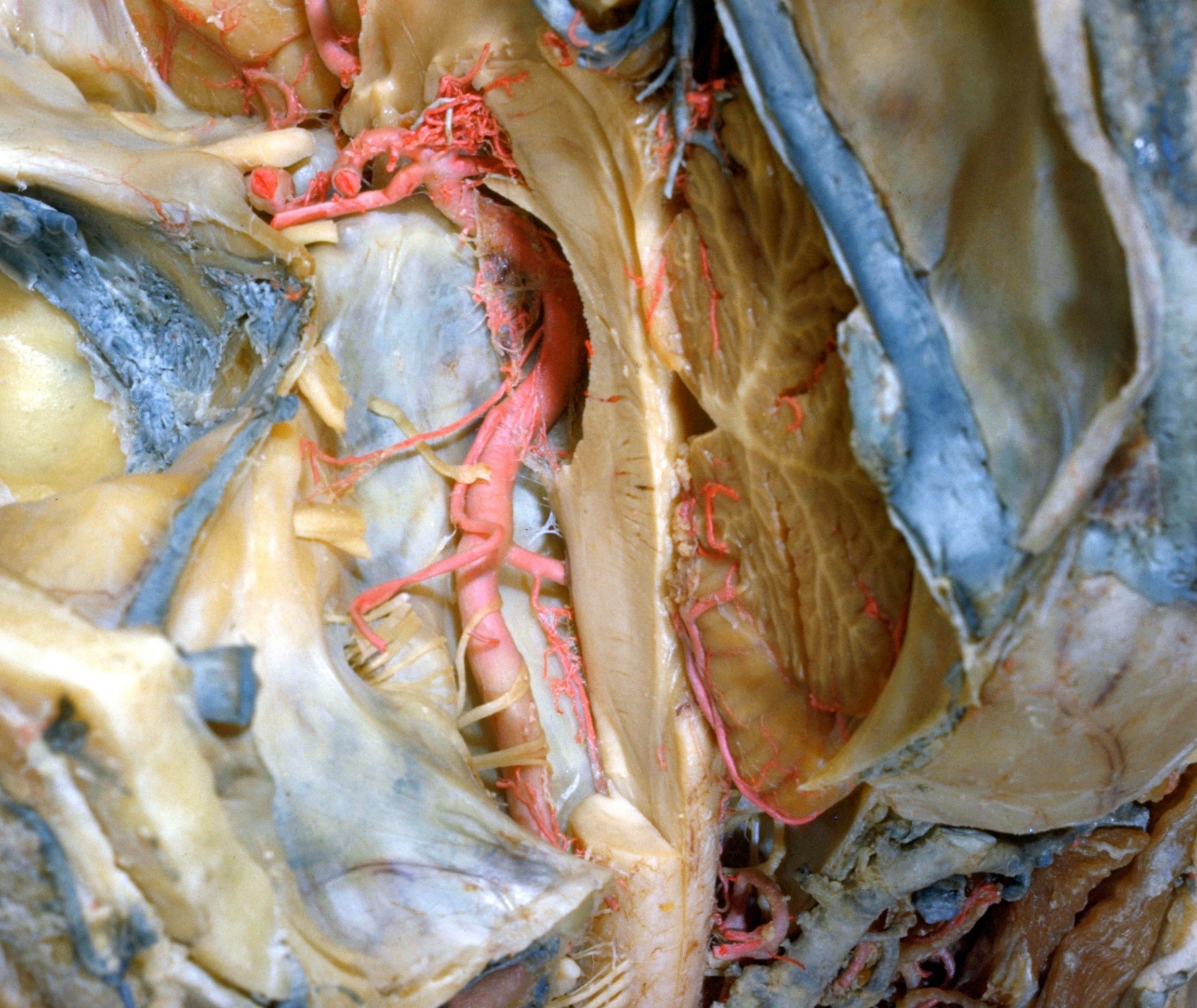
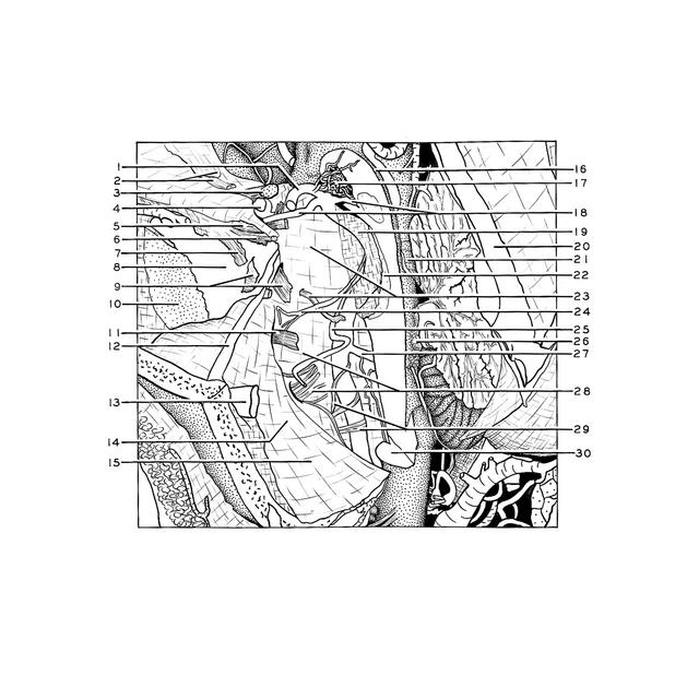
Exploration of the meninges and brain in situ
Posterior cranial fossa, basilar and vertebral arteries, petrosal sinuses and basilar plexus of veins
Stanford holds the copyright to the David L. Bassett anatomical images and has assigned
Creative Commons license Attribution-Share
Alike 4.0 International to all of the images.
For additional information regarding use and permissions,
please contact the Medical History Center.
Image #4-3
Exploration of the meninges and brain in situ
Posterior cranial fossa, basilar and vertebral arteries, petrosal sinuses and basilar plexus of veins
The left half of the brain stem has been removed and the basilar part of the pons cut away on the right to reveal the course of the basilar artery (19). The inferior petrosal sinus and basilar plexus have not been opened but their positions under the dura are identified by the blue color of the latex with which they are filled.
- Posterior cerebral artery left (cut across)
- Falx cerebri (upper pointer) and olfactory tract
- Optic nerve (II)
- Internal carotid artery left (cut across)
- Oculomotor nerve (III) within cavernous sinus
- Trochlear nerve (IV) within cavernous sinus
- Ophthalmic nerve (VI) within cavernous sinus
- Cavernous sinus
- Semilunar ganglion (trigeminal) and trigeminal nerve (V)
- Middle cranial fossa (dura removed)
- Facial nerve (VII) and Vestibulocochlear (VIII) entering internal acoustic meatus
- Superior petrosal sinus (opened)
- Transverse sinus
- Position of sigmoid portion of transverse sinus (unopened)
- Posterior cranial fossa
- Cerebral aqueduct
- Arteries within interpeduncular fossa
- Superior cerebellar arteries
- Basilar artery
- Straight sinus
- Cerebellar lingula
- Basilar part of pons (cut away)
- Basilar plexus (upper pointer) and abducens nerve (VI)
- Internal auditory artery
- Anterior inferior cerebellar artery
- Choroid plexus fourth ventricle
- Vertebral artery right
- Vagus nerve (X) and inferior petrosal sinus (upper pointer indicates position of this sinus beneath the dura)
- Roots hypoglossal nerve (XII)
- Medulla oblongata (cut across to midline)