Exploration of the meninges and brain in situ
Middle cranial fossa, middle meningeal vessels and cavernous sinus
Stanford holds the copyright to the David L. Bassett anatomical images and has assigned
Creative Commons license Attribution-Share
Alike 4.0 International to all of the images.
For additional information regarding use and permissions,
please contact the Medical History Center.
Image #4-2
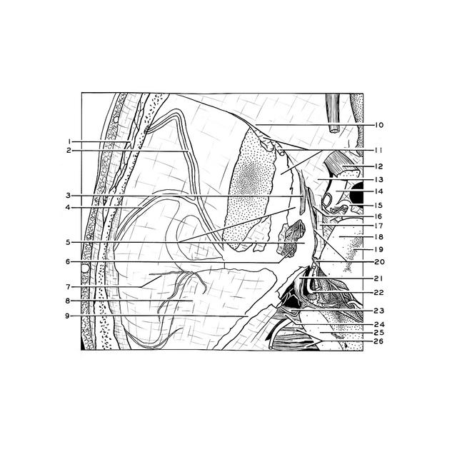
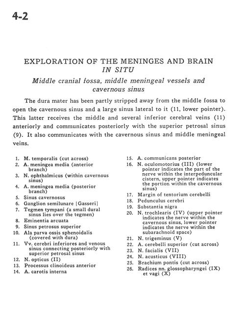
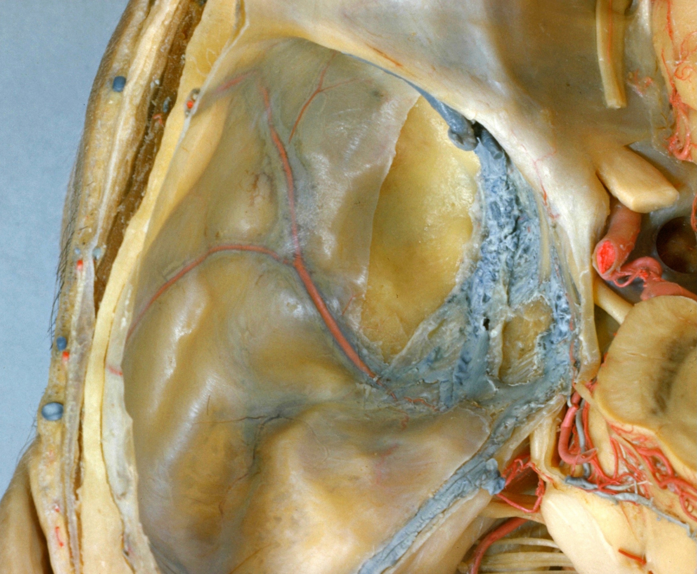
Exploration of the meninges and brain in situ
Middle cranial fossa, middle meningeal vessels and cavernous sinus
The dura mater has been partly stripped away from the middle fossa to open the cavernous sinus and a large sinus lateral to it (11, lower pointer). This latter receives the middle and several inferior cerebral veins (11) anteriorly and communicates posteriorly with the superior petrosal sinus (9). It also communicates with the cavernous sinus and middle meningeal veins.
- Temporalis muscle (cut across)
- Middle meningeal artery (anterior branch)
- Ophthalmic nerve (VI) (within cavernous sinus)
- Middle meningeal artery (posterior branch)
- Cavernous sinus
- Semilunar ganglion (trigeminal)
- Tegmen tympani (a small dural sinus lies over the tegmen)
- Arcuate eminence
- Superior petrosal sinus
- Lesser wing sphenoid bone (covered with dura)
- Inferior cerebral vein and venous sinus connecting posteriorly with superior petrosal sinus
- Optic nerve (II)
- Anterior clinoid process
- Internal carotid artery
- Posterior communicating artery
- Oculomotor nerve (III) (lower pointer indicates the part of the nerve within the interpeduncular cistern, upper pointer indicates the portion within the cavernous sinus)
- Margin of tentorium cerebelli
- Cerebral peduncle
- Substantia nigra
- Trochlear nerve (IV) (upper pointer indicates the nerve within the cavernous sinus, lower pointer indicates the nerve within the subarachnoid space)
- Trigeminal nerve (V)
- Superior cerebellar artery (cut across)
- Facial nerve (VII)
- Vestibulocochlear nerve (VIII)
- Brachium pontis (middle cerebellar peduncle) (cut across)
- Roots glossopharyngeal nerve (IX) and vagus (X)