Osteology
Lateral wall of left nasal fossa dissected
Stanford holds the copyright to the David L. Bassett anatomical images and has assigned
Creative Commons license Attribution-Share
Alike 4.0 International to all of the images.
For additional information regarding use and permissions,
please contact the Medical History Center.
Image #38-2
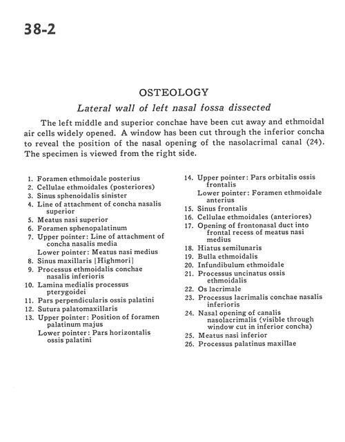
Osteology
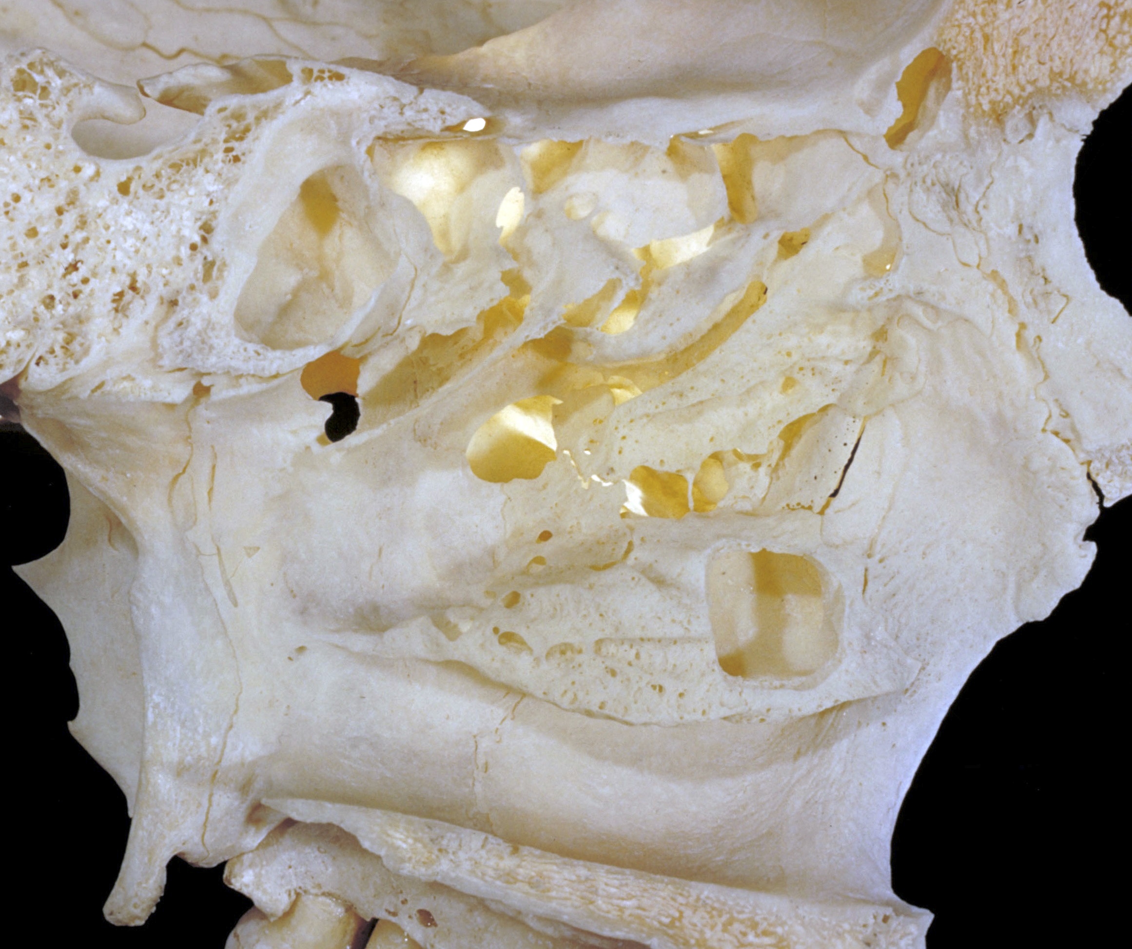
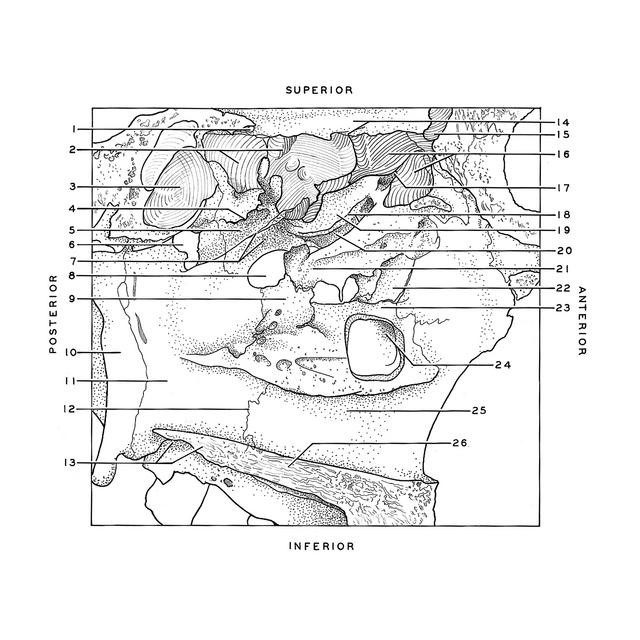
Lateral wall of left nasal fossa dissected
The left middle and superior conchae have been cut away and ethmoidal air cells widely opened. A window has been cut through the inferior concha to reveal the position of the nasal opening of the nasolacrimal canal (24). The specimen is viewed from the right side.
- Posterior ethmoidal foramen
- Ethmoidal cells (posterior)
- Sphenoid sinus left
- Line of attachment of superior nasal concha
- Superior nasal meatus
- Sphenopalatine foramen
- Upper pointer: Line of attachment of middle nasal concha Lower pointer: Middle nasal meatus
- Maxillary sinus
- Ethmoidal process inferior nasal conchae
- Medial plate of pterygoid process
- Perpendicular part palatine bone
- Palatomaxillary suture
- Upper pointer: Position of greater palatine foramen Lower pointer: Horizontal plate palatine bone
- Upper pointer: Orbital part of frontal bone Lower pointer: Anterior ethmoidal foramen
- Frontal sinus
- Ethmoidal cells (anterior)
- Opening of frontonasal duct into frontal recess of middle nasal meatus
- Hiatus semilunaris
- Ethmoidal bulla
- Ethmoidal infundibulum
- Uncinate process ethmoid bone
- Lacrimal bone
- Lacrimal process inferior nasal conchae
- Nasal opening of nasolacrimal canal visible through window cut in inferior concha)
- Inferior nasal meatus
- Palatine process of maxilla