
Radiographs of the brain
Lateral pneumoencephalogram; lateral ventricles and third ventricle
Stanford holds the copyright to the David L. Bassett anatomical images and has assigned
Creative Commons license Attribution-Share
Alike 4.0 International to all of the images.
For additional information regarding use and permissions,
please contact the Medical History Center.
Image #34-5
Radiographs of the brain
Lateral pneumoencephalogram; lateral ventricles and third ventricle
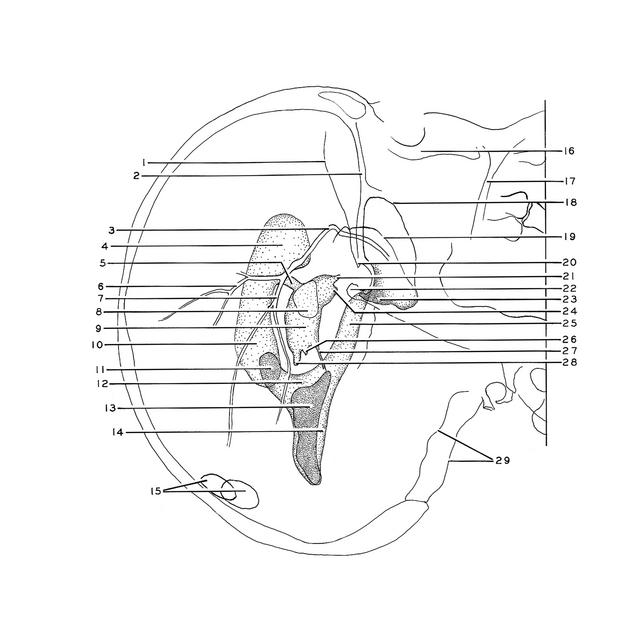
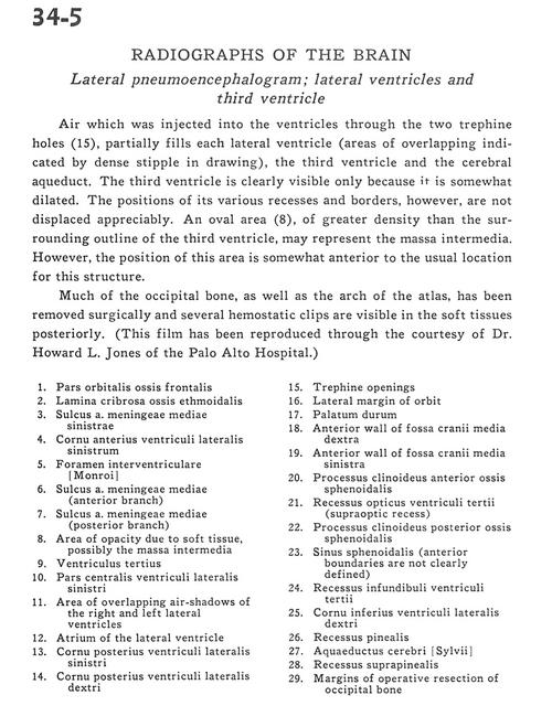
Air which was injected into the ventricles through the two trephine holes (15), partially fills each lateral ventricle (areas of overlapping indicated by dense stipple in drawing), the third ventricle and the cerebral aqueduct. The third ventricle is clearly visible only because it is somewhat dilated. The positions of its various recesses and borders, however, are not displaced appreciably. An oval area (8), of greater density than the surrounding outline of the third ventricle, may represent the massa intermedia. However, the position of this area is somewhat anterior to the usual location for this structure.
- Orbital part of frontal bone
- Cribriform plate ethmoid bone
- Sulcus of left middle meningeal artery
- Anterior horn lateral ventricle left
- Interventricular foramen
- Sulcus of middle meningeal artery (anterior branch)
- Sulcus middle meningeal artery (posterior branch)
- Area of opacity due to soft tissue, possibly the massa intermedia
- Third ventricle
- Central part of left lateral ventricle
- Area of overlapping air shadows of the right and left lateral ventricles
- Atrium of the lateral ventricle
- Posterior horn lateral ventricle left
- Posterior horn lateral ventricle right
- Trephine openings
- Lateral margin of orbit
- Hard palate
- Anterior wall of middle cranial fossa right
- Anterior wall of middle cranial fossa left
- Anterior clinoid process sphenoid bone
- Optic recess third ventricle (supraoptic recess)
- Posterior clinoid process sphenoid bone
- Sphenoidal sinus (anterior boundaries are not clearly defined)
- Infundibular recess third ventricle
- Inferior horn of lateral ventricle right
- Pineal recess
- Cerebral aqueduct
- Suprapineal recess
- Margins of operative resection of occipital bone