Radiographs of the brain
Right vertebral angiogram, arterial phase
Stanford holds the copyright to the David L. Bassett anatomical images and has assigned
Creative Commons license Attribution-Share
Alike 4.0 International to all of the images.
For additional information regarding use and permissions,
please contact the Medical History Center.
Image #34-3
Radiographs of the brain
Right vertebral angiogram, arterial phase
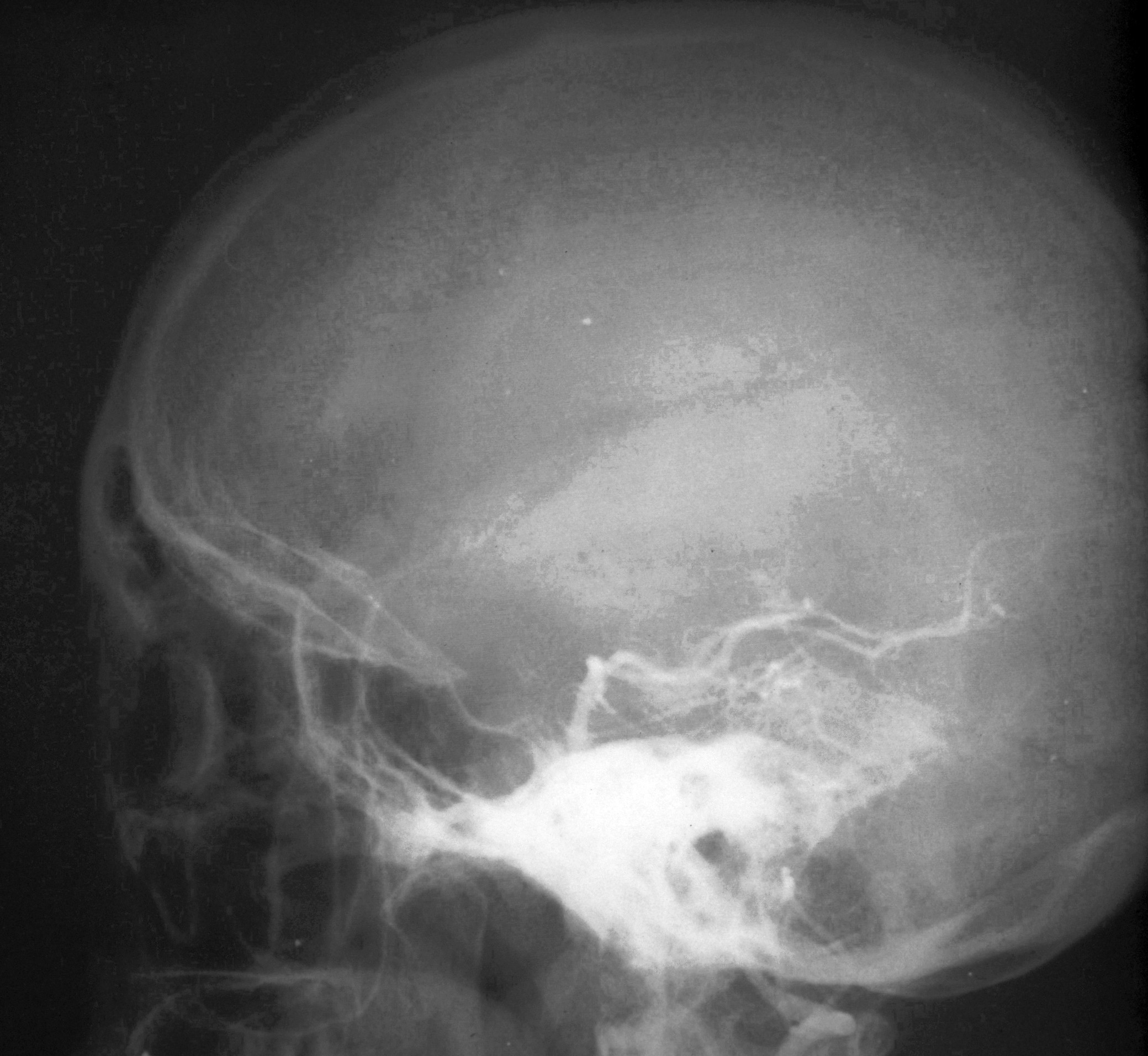
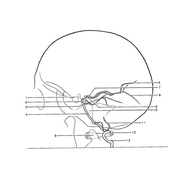
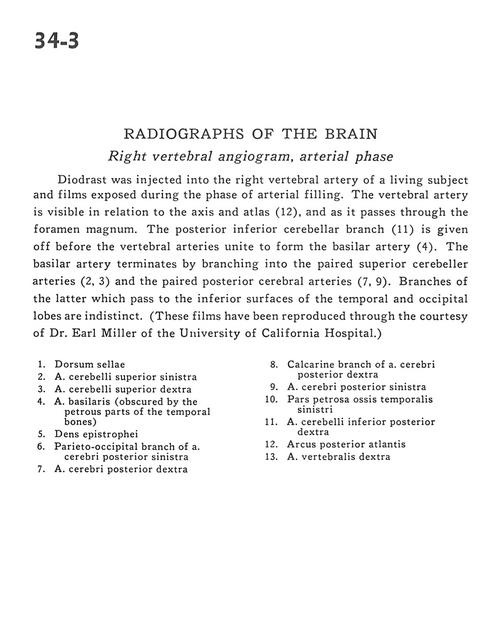
Diodrast was injected into the right vertebral artery of a living subject and films exposed during the phase of arterial filling. The vertebral artery is visible in relation to the axis and atlas (12), and as it passes through the foramen magnum. The posterior inferior cerebellar branch (11) is given off before the vertebral arteries unite to form the basilar artery (4). The basilar artery terminates by branching into the paired superior cerebellar arteries (2, 3) and the paired posterior cerebral arteries (7, 9). Branches of the latter which pass to the inferior surfaces of the temporal and occipital lobes are indistinct. (These films have been reproduced through the courtesy of Dr. Earl Miller of the University of California Hospital.)
- Dorsum sellae
- Superior cerebellar artery left
- Superior cerebellar artery right
- Basilar artery (obscured by the petrous parts of the temporal bones)
- Dens
- Parieto-occipital branch of posterior cerebral artery left
- Posterior cerebral artery right
- Calcarine branch of posterior cerebral artery right
- Posterior cerebral artery left
- Petrosal part of temporal bone left
- Posterior inferior cerebellar artery right
- Posterior arch of atlas
- Vertebral artery right