Radiographs of the brain
Left internal carotid angiogram, arterial phase
Stanford holds the copyright to the David L. Bassett anatomical images and has assigned
Creative Commons license Attribution-Share
Alike 4.0 International to all of the images.
For additional information regarding use and permissions,
please contact the Medical History Center.
Image #34-1
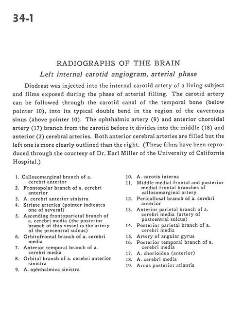
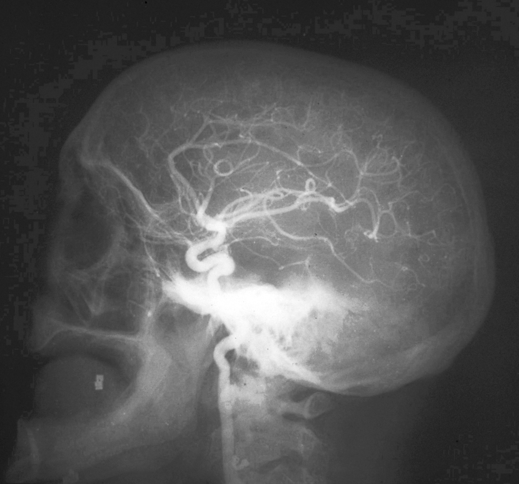
Radiographs of the brain
Left internal carotid angiogram, arterial phase
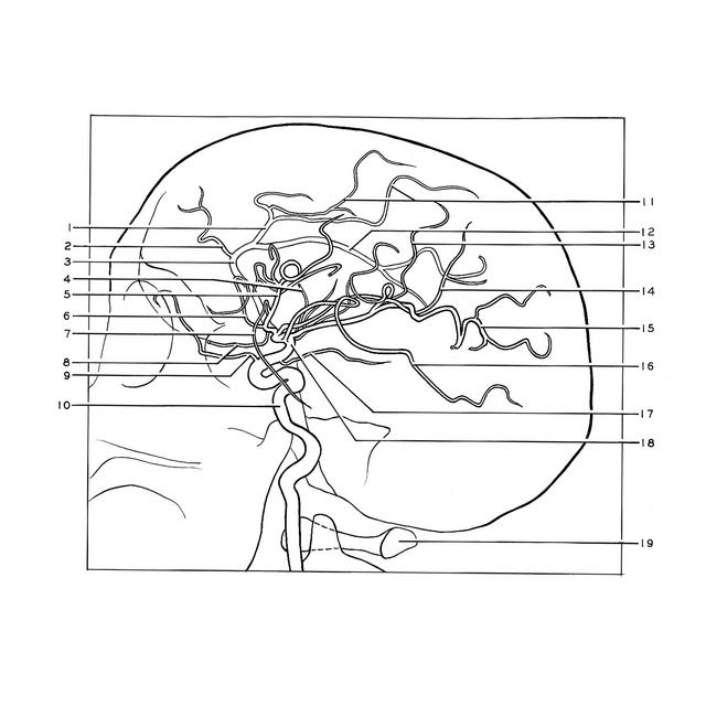
Diodrast was injected into the internal carotid artery of a living subject and films exposed during the phase of arterial filling. The carotid artery can be followed through the carotid canal of the temporal bone (below pointer 10), into its typical double bend in the region of the cavernous sinus (above pointer 10). The ophthalmic artery (9) and anterior choroidal artery (17) branch from the carotid before it divides into the middle (18) and anterior (3) cerebral arteries. Both anterior cerebral arteries are filled but the left one is more clearly outlined than the right. (These films have been reproduced through the courtesy of Dr. Earl Miller of the University of California Hospital.)
- Callosomarginal branch of anterior cerebral artery
- Frontopolar branch of anterior cerebral artery
- Anterior cerebral artery left
- Striate arteries (pointer indicates one of several)
- Ascending frontoparietal branch of middle cerebral artery (the posterior branch of this vessel is the artery of the precentral sulcus)
- Orbitofrontal branch of middle cerebral artery
- Anterior temporal branch of middle cerebral artery
- Orbital branch of anterior cerebral artery left
- Ophthalmic artery left
- Internal carotid artery
- Middle medial frontal and posterior medial frontal branches of callosomarginal artery
- Pericallosal branch of anterior cerebral artery
- Anterior parietal branch of middle cerebral artery (artery of postcentral sulcus)
- Posterior parietal branch of middle cerebral artery
- Artery of angular gyrus
- Posterior temporal branch of middle cerebral artery
- Choroidal artery (anterior)
- Middle cerebral artery
- Posterior arch of atlas