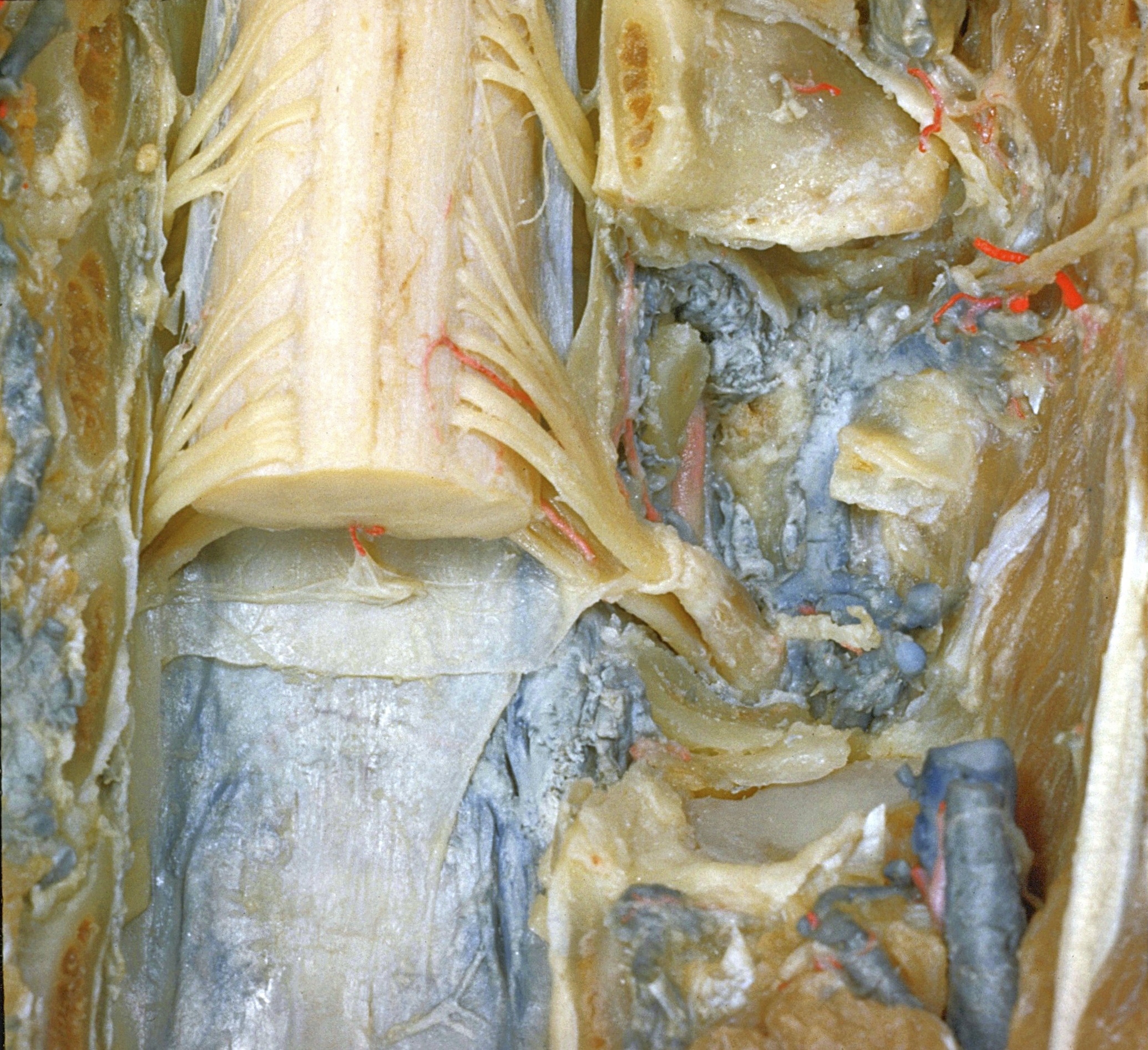
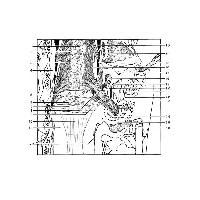
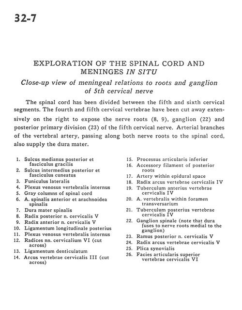
Exploration of the spinal cord and meninges in situ
Close-up view of meningeal relations to roots and ganglion of 5th cervical nerve
Stanford holds the copyright to the David L. Bassett anatomical images and has assigned
Creative Commons license Attribution-Share
Alike 4.0 International to all of the images.
For additional information regarding use and permissions,
please contact the Medical History Center.
Image #32-7
Exploration of the spinal cord and meninges in situ
Close-up view of meningeal relations to roots and ganglion of 5th cervical nerve
The spinal cord has been divided between the fifth and sixth cervical segments. The fourth and fifth cervical vertebrae have been cut away extensively on the right to expose the nerve roots (8, 9), ganglion (22) and posterior primary division (23) of the fifth cervical nerve. Arterial branches of the vertebral artery, passing along both nerve roots to the spinal cord, also supply the dura mater.
- Posterior median sulcus and gracile fasciculus
- Posterior intermediate sulcus and cuneate fasciculus
- Lateral funiculus
- Internal vertebral venous plexus
- Gray columns of spinal cord
- Anterior spinal artery and arachnoid
- Dura mater
- Dorsal root cervical nerve V
- Ventral root cervical nerve V
- Posterior longitudinal ligament
- Internal vertebral venous plexus
- Roots cervical nerve VI (cut across)
- Denticulate ligament
- Arch of cervical vertebra IV (cut across)
- Inferior articular process
- Accessory filament of posterior roots
- Artery within epidural space
- Root arch of cervical vertebra V
- Anterior tubercle cervical vertebra V
- Vertebral artery within transverse foramen
- Posterior tubercle cervical vertebra V
- Spinal ganglion (note that dura fuses to nerve roots medial to the ganglion)
- Posterior branch of cervical nerve V
- Root arch of cervical vertebra VI
- Synovial fold
- Superior articular surface of cervical vertebra VII