Exploration of the spinal cord and meninges in situ
Cervical spinal cord; denticulate ligaments
Stanford holds the copyright to the David L. Bassett anatomical images and has assigned
Creative Commons license Attribution-Share
Alike 4.0 International to all of the images.
For additional information regarding use and permissions,
please contact the Medical History Center.
Image #32-6
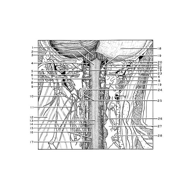
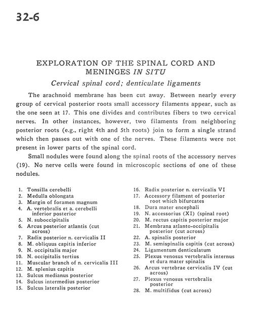
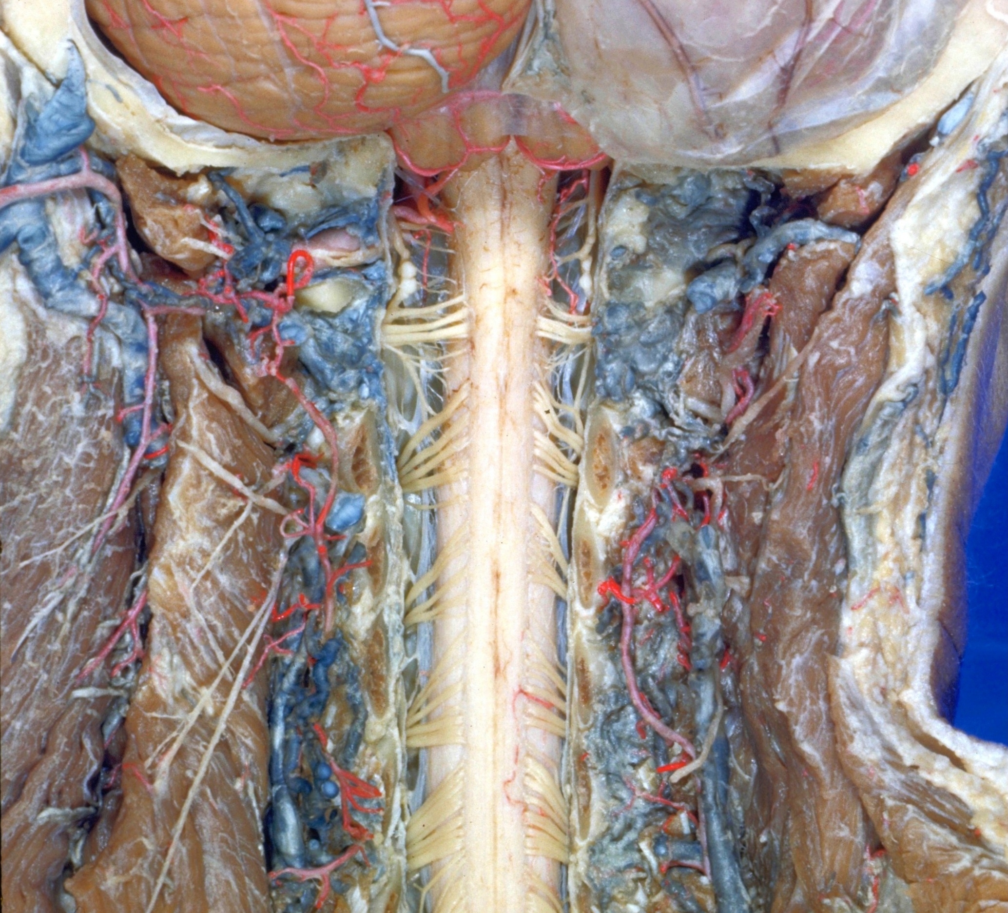
Exploration of the spinal cord and meninges in situ
Cervical spinal cord; denticulate ligaments
The arachnoid membrane has been cut away. Between nearly every group of cervical posterior roots small accessory filaments appear, such as the one seen at 17. This one divides and contributes fibers to two cervical nerves. In other instances, however, two filaments from neighboring posterior roots (e.g., right 4th and 5th roots) join to form a single strand which then passes out with one of the nerves. These filaments were not present in lower parts of the spinal cord.
- Cerebellar tonsil
- Medulla oblongata
- Margin of foramen magnum
- Vertebral artery and posterior inferior cerebellar artery
- Suboccipital nerve
- Posterior arch of atlas (cut across)
- Dorsal root cervical nerve II
- Inferior oblique capitis muscle
- Greater occipital nerve
- Third occipital nerve
- Muscular branch of cervical nerve III
- Splenius capitis muscle
- Posterior median sulcus
- Posterior intermedian sulcus
- Posterior lateral sulcus
- Dorsal root cervical nerve VI
- Accessory filament of posterior root which bifurcates
- Dura mater
- Accessory nerve (Xl) (spinal root)
- Posterior major rectus capitis muscle
- Posterior atlanto-occipital membrane (cut across)
- Posterior spinal artery
- Semispinalis capitis muscle (cut across)
- Denticulate ligament
- Internal vertebral venous plexus and dura mater
- Arch of cervical vertebra IV (cut across)
- Posterior vertebral venous plexus
- Multifidus muscle (cut across)