Serial transverse sections of the brain stem
Junction of mesencephalon and diencephalon.
Stanford holds the copyright to the David L. Bassett anatomical images and has assigned
Creative Commons license Attribution-Share
Alike 4.0 International to all of the images.
For additional information regarding use and permissions,
please contact the Medical History Center.
Image #31-1
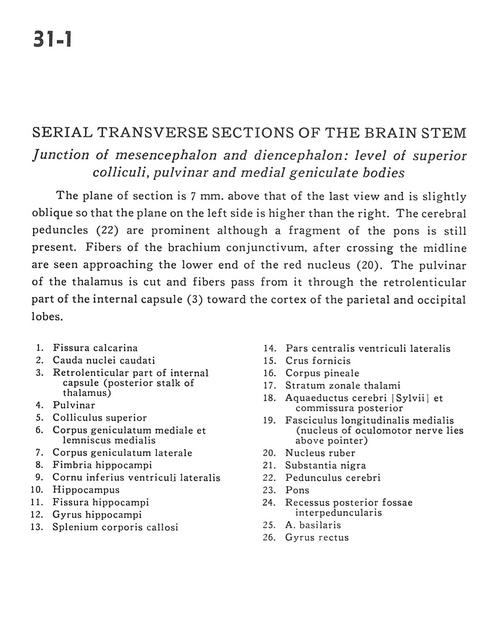
Serial transverse sections of the brain stem
Junction of mesencephalon and diencephalon.
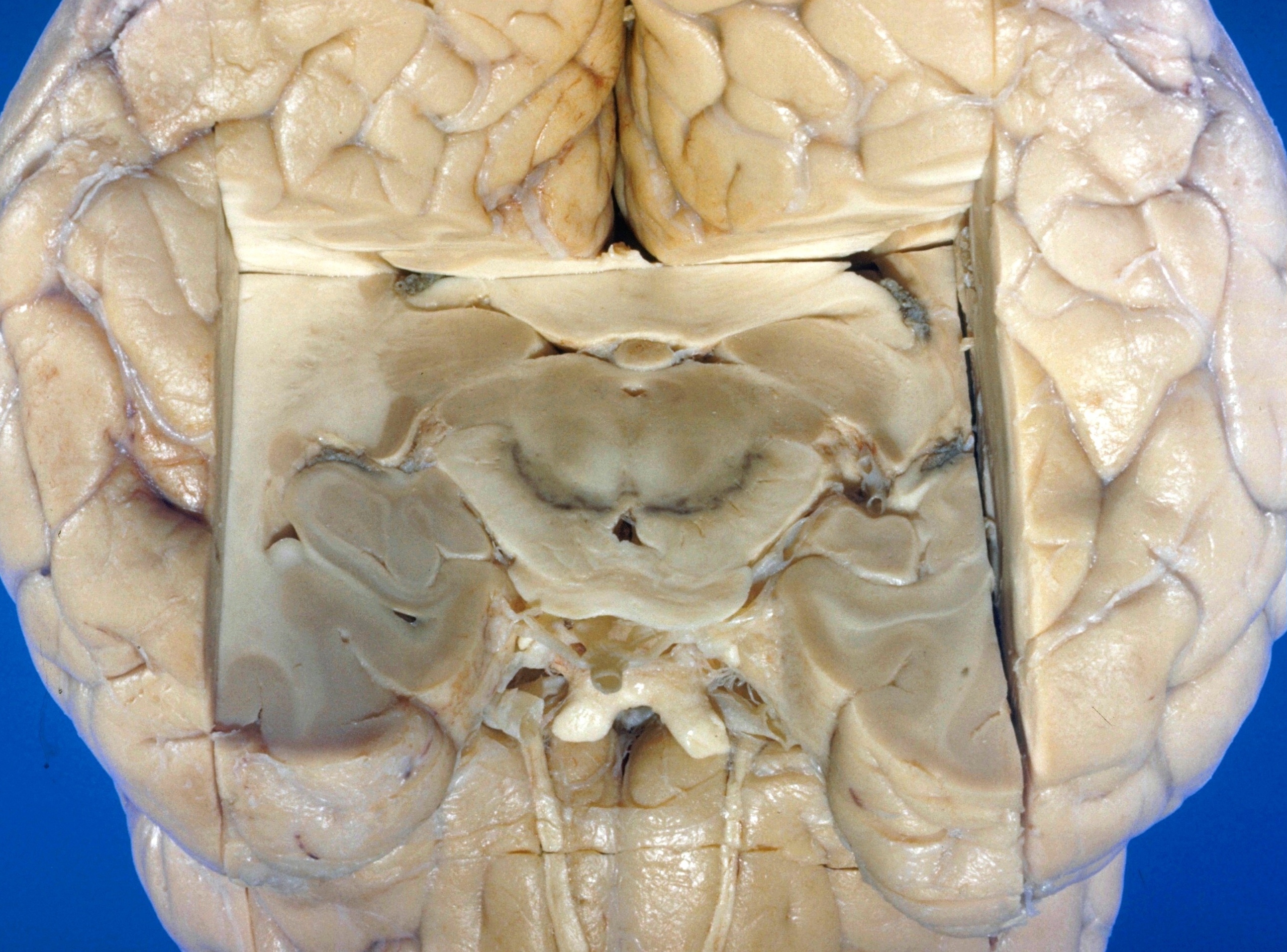
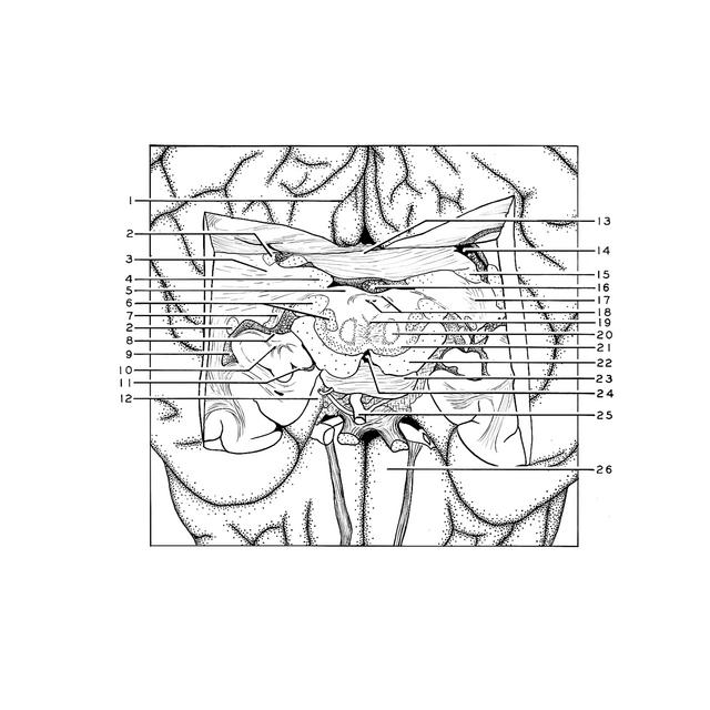
The plane of section is 7 mm. above that of the last view and is slightly oblique so that the plane on the left side is higher than the right. The cerebral peduncles (22) are prominent although a fragment of the pons is still present. Fibers of the brachium conjunctivum, after crossing the midline are seen approaching the lower end of the red nucleus (20). The pulvinar of the thalamus is cut and fibers pass from it through the retrolenticular part of the internal capsule (3) toward the cortex of the parietal and occipital lobes.
- Calcarine fissure
- Caudate nucleus (tail)
- Retrolenticular part of internal capsule (posterior stalk of thalamus)
- Pulvinar
- Superior colliculus
- Medial geniculate body and medial lemniscus
- Lateral geniculate body
- Hippocampal fimbria
- Inferior horn of lateral ventricle
- Hippocampus
- Hippocampal fissure
- Hippocampal gyrus
- Corpus callosum (splenium)
- Central part lateral ventricle
- Fornix (crus)
- Pineal body
- Stratum zonale thalami
- Cerebral aqueduct and posterior commissure
- Medial longitudinal fasciculus (nucleus of oculomotor nerve lies above pointer)
- Red nucleus
- Substantia nigra
- Cerebral peduncle
- Pons
- Posterior recess of interpeduncular fossa
- Basilar artery
- Straight gyrus