Exploration of the meninges and brain in situ
Close-up view of diaphragma sellae and infundibulum
Stanford holds the copyright to the David L. Bassett anatomical images and has assigned
Creative Commons license Attribution-Share
Alike 4.0 International to all of the images.
For additional information regarding use and permissions,
please contact the Medical History Center.
Image #3-4
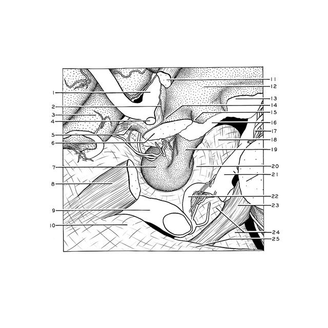
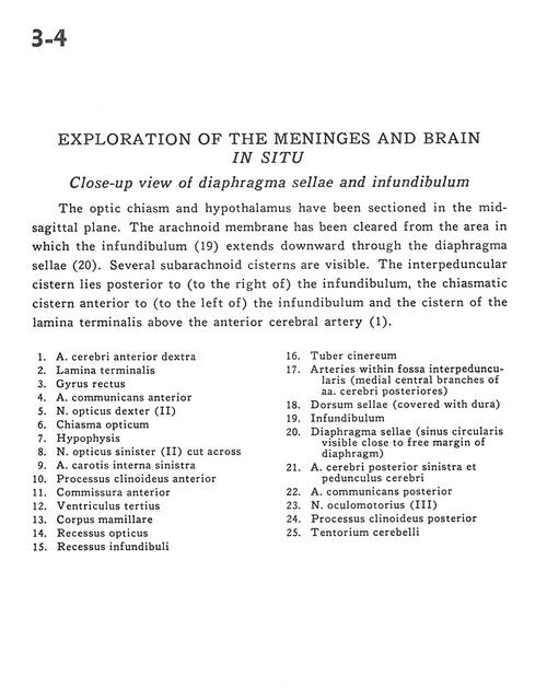
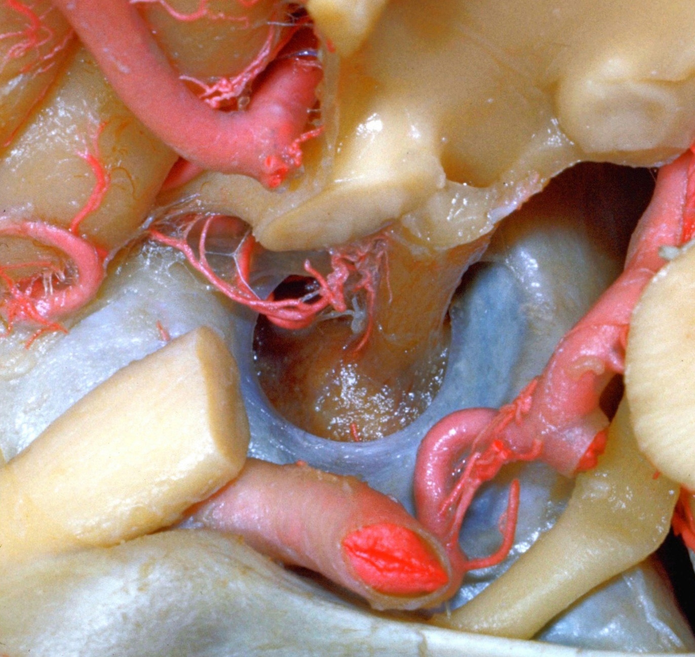
Exploration of the meninges and brain in situ
Close-up view of diaphragma sellae and infundibulum
The optic chiasm and hypothalamus have been sectioned in the mid-sagittal plane. The arachnoid membrane has been cleared from the area in which the infundibulum (19) extends downward through the diaphragma sellae (20). Several subarachnoid cisterns are visible. The interpeduncular cistern lies posterior to (to the right of) the infundibulum, the chiasmatic cistern anterior to (to the left of) the infundibulum and the cistern of the lamina terminalis above the anterior cerebral artery (1).
- Anterior cerebral artery right
- Lamina terminalis
- Gyrus rectus
- Anterior communicating artery
- Right optic nerve (II)
- Optic chiasm
- Hypophysis
- Left optic nerve (II) (cut across)
- Internal carotid artery left
- Anterior clinoid process
- Anterior commissure
- Third ventricle
- Mamillary body
- Optic recess
- Infundibular recess
- Tuber cinereum
- Arteries within interpeduncular fossa (medial central branches of posterior cerebral artery)
- Dorsum sellae (covered with dura)
- Infundibulum
- Diaphragma sellae (sinus circularis visible close to free margin of diaphragm)
- Posterior cerebral artery left and cerebral peduncle
- Posterior communicating artery
- Oculomotor nerve (III)
- Posterior clinoid process
- Tentorium cerebelli