Serial transverse sections of the brain stem
Medulla oblongata.
Stanford holds the copyright to the David L. Bassett anatomical images and has assigned
Creative Commons license Attribution-Share
Alike 4.0 International to all of the images.
For additional information regarding use and permissions,
please contact the Medical History Center.
Image #29-7
Serial transverse sections of the brain stem
Medulla oblongata.
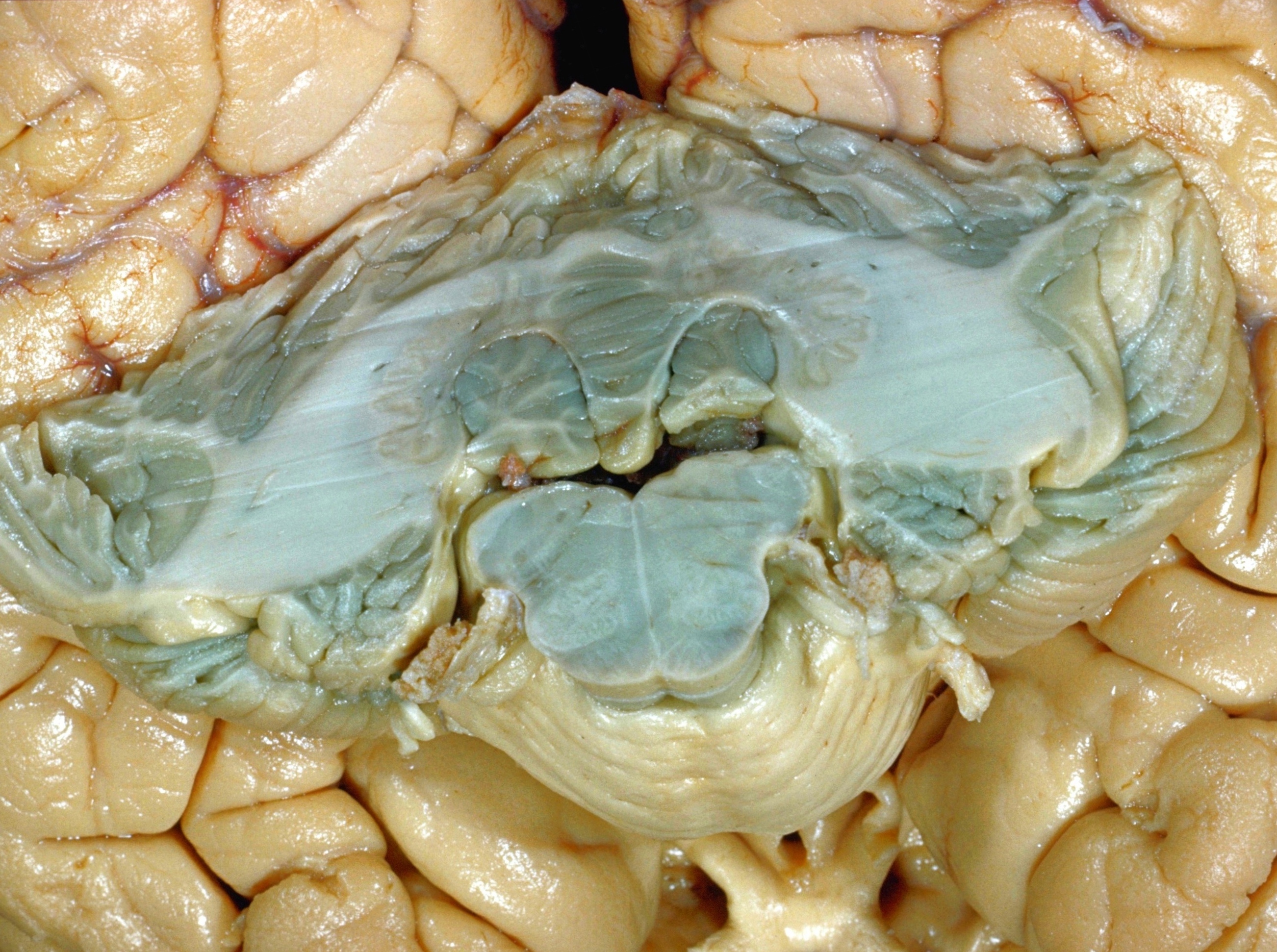
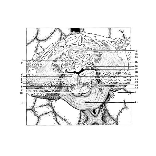
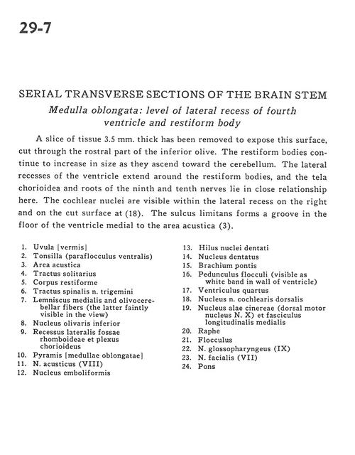
A slice of tissue 3.5 mm. thick has been removed to expose this surface, cut through the rostral part of the inferior olive. The restiform bodies continue to increase in size as they ascend toward the cerebellum. The lateral recesses of the ventricle extend around the restiform bodies, and the tela chorioidea and roots of the ninth and tenth nerves lie in close relationship here. The cochlear nuclei are visible within the lateral recess on the right and on the cut surface at (18). The sulcus limitans forms a groove in the floor of the ventricle medial to the area acustica (3).
- Uvula (vermis)
- Tonsil (ventral paraflocculus)
- Area acustica
- Tractus solitarius
- Restiform body (inferior cerebellar peduncle)
- Spinal trigeminal tract
- Medial lemniscus and olivocerebellar fibers (the latter faintly visible in the view)
- Inferior olivary nucleus
- Lateral recess of rhomboid fossa and choroid plexus
- Pyramid (medulla oblongata)
- Vestibulocochlear nerve (VIII)
- Emboliform nucleus
- Hilus dentate nucleus
- Dentate nucleus
- Brachium pontis (middle cerebellar peduncle)
- Floccular peduncle (visible as white band in wall of ventricle)
- Fourth ventricle
- Dorsal cochlear nucleus
- Dorsal motor nucleus of vagus nerve (X) and medial longitudinal fasciculus
- Raphe
- Flocculus
- Glossopharyngeal nerve (IX)
- Facial nerve (VII)
- Pons