Serial transverse sections of the brain stem
Medulla oblongata.
Stanford holds the copyright to the David L. Bassett anatomical images and has assigned
Creative Commons license Attribution-Share
Alike 4.0 International to all of the images.
For additional information regarding use and permissions,
please contact the Medical History Center.
Image #29-3
Serial transverse sections of the brain stem
Medulla oblongata.
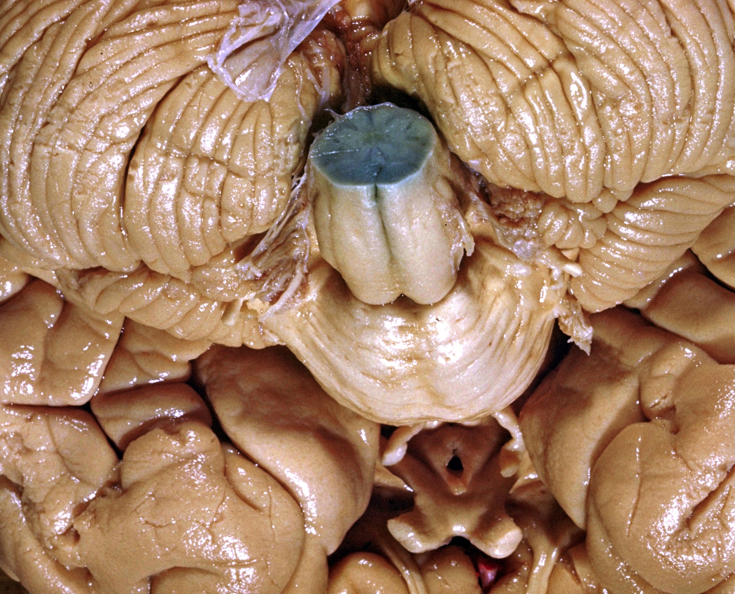
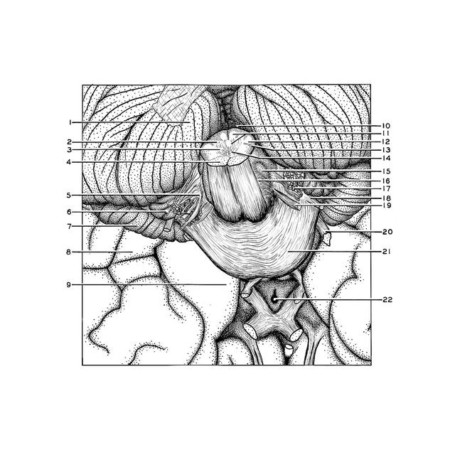
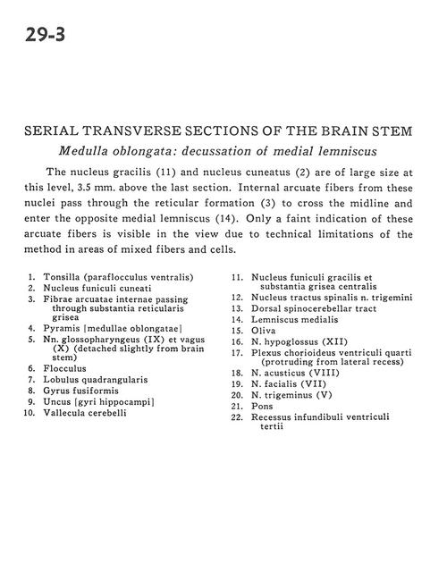
The nucleus gracilis (11) and nucleus cuneatus (2) are of large size at this level, 3.5 mm. above the last section. Internal arcuate fibers from these nuclei pass through the reticular formation (3) to cross the midline and enter the opposite medial lemniscus (14). Only a faint indication of these arcuate fibers is visible in the view due to technical limitations of the method in areas of mixed fibers and cells.
- Tonsil (ventral paraflocculus)
- Cuneate nucleus
- Internal arcuate fibers passing through reticular gray matter
- Pyramid (medulla oblongata)
- Glossopharyngeal nerve (IX) and vagus (X) (detached slightly from brain stem)
- Flocculus
- Quadrangular lobule
- Fusiform gyrus
- Uncus (hippocampal gyrus)
- Vallecula cerebelli
- Gracile nucleus and central gray matter
- Spinal trigeminal nucleus
- Dorsal spinocerebellar tract
- Medial lemniscus
- Olive
- Hypoglossal nerve (XII)
- Choroid plexus fourth ventricle (protruding from lateral recess)
- Vestibulocochlear nerve (VIII)
- Facial nerve (VII)
- Trigeminal nerve (V)
- Pons
- Infundibular recess third ventricle