Exploration of the basal aspects of the medulla, pons and cerebellum
Spinal tract of trigeminal nerve
Stanford holds the copyright to the David L. Bassett anatomical images and has assigned
Creative Commons license Attribution-Share
Alike 4.0 International to all of the images.
For additional information regarding use and permissions,
please contact the Medical History Center.
Image #28-7
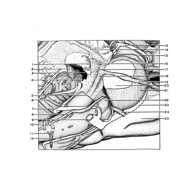
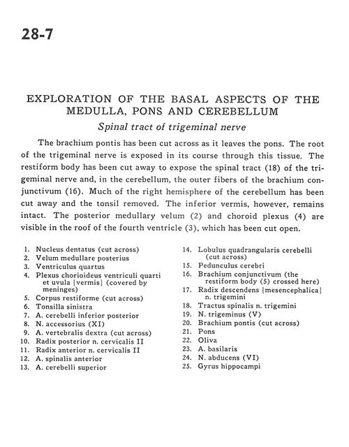
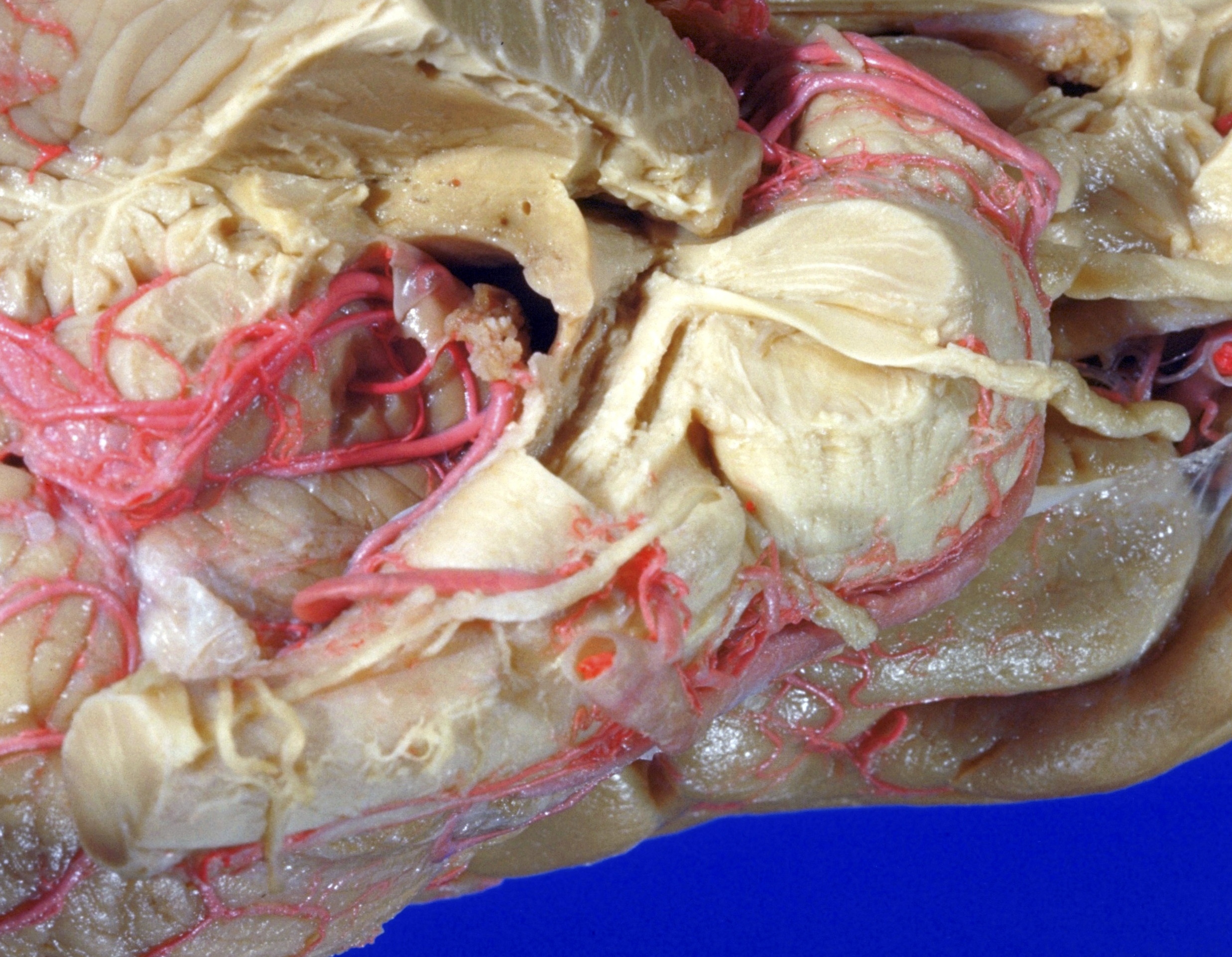
Exploration of the basal aspects of the medulla, pons and cerebellum
Spinal tract of trigeminal nerve
The brachium pontis has been cut across as it leaves the pons. The root of the trigeminal nerve is exposed in its course through this tissue. The restiform body has been cut away to expose the spinal tract (18) of the trigeminal nerve and, in the cerebellum, the outer fibers of the brachium conjunctivum (16). Much of the right hemisphere of the cerebellum has been cut away and the tonsil removed. The inferior vermis, however, remains intact. The posterior medullary velum (2), and choroid plexus (4) are visible in the roof of the fourth ventricle (3), which has been cut open.
- Dentate nucleus (cut across)
- Posterior medullary velum
- Fourth ventricle
- Choroid plexus fourth ventricle and uvula (covered by meninges)
- Restiform body (inferior cerebellar peduncle) (cut across)
- Tonsil left
- Posterior inferior cerebellar artery
- Accessory nerve (XI)
- Vertebral artery right (cut across)
- Dorsal root cervical nerve II
- Ventral root cervical nerve II
- Anterior spinal artery
- Superior cerebellar artery
- Quadrangular lobule of cerebellum (cut across)
- Cerebral peduncle
- Brachium conjunctivum (the restiform body (5) crossed here)
- Descending root (mesencephalic) of trigeminal nerve (V)
- Spinal trigeminal tract
- Trigeminal nerve (V)
- Brachium pontis (cut across)
- Pons
- Olive
- Basilar artery
- Abducens nerve (VI)
- Hippocampal gyrus