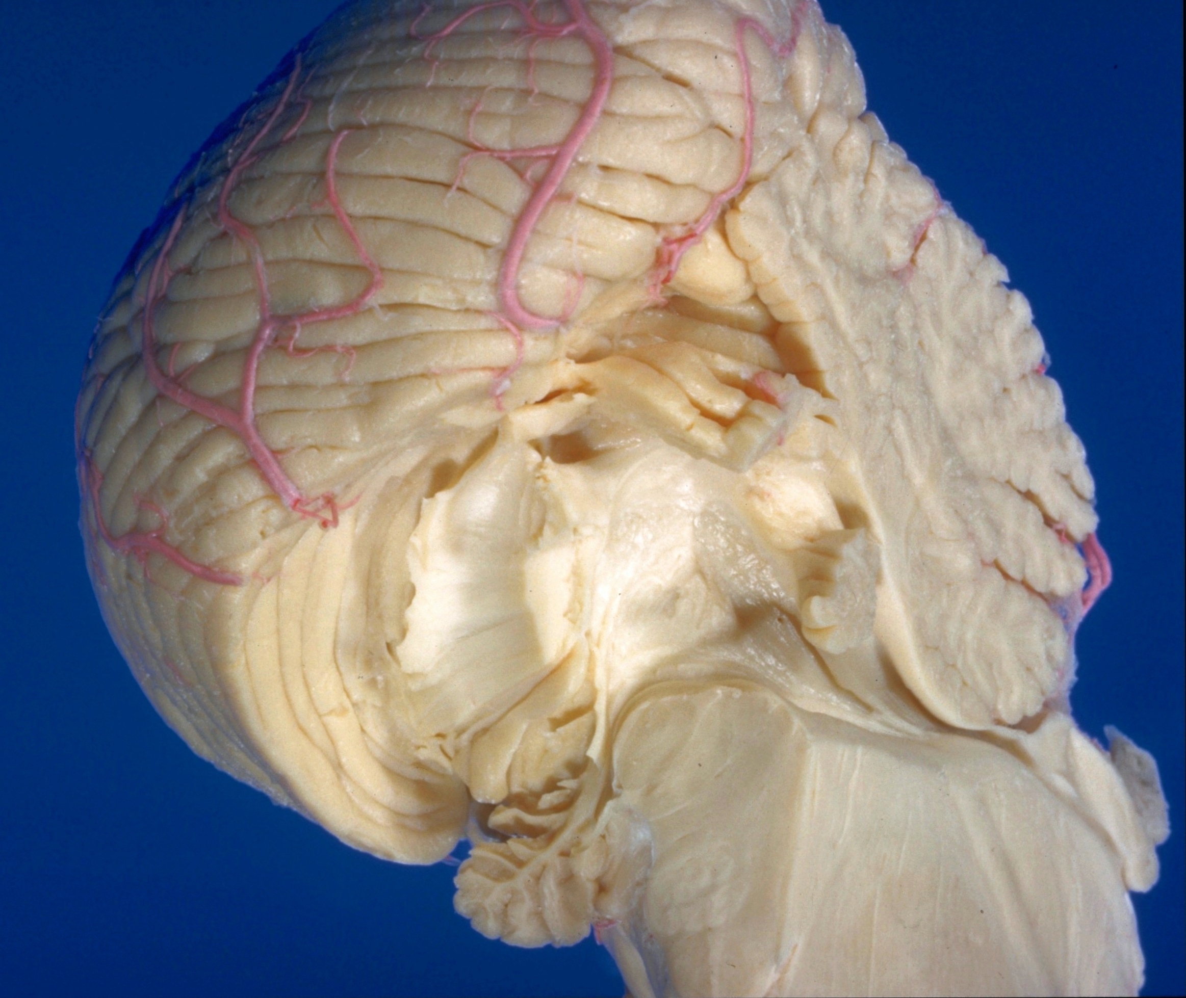
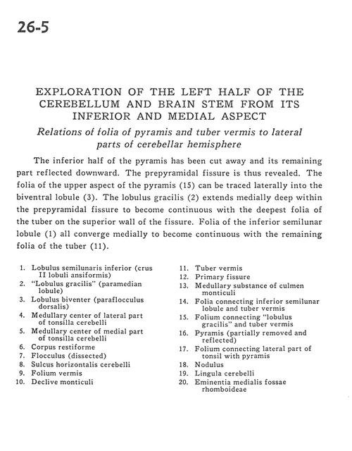
Exploration of the left half of the cerebellum and brain stem from its inferior and medial aspect
Relations of folia of pyramis and tuber vermis to lateral parts of cerebellar hemisphere
Stanford holds the copyright to the David L. Bassett anatomical images and has assigned
Creative Commons license Attribution-Share
Alike 4.0 International to all of the images.
For additional information regarding use and permissions,
please contact the Medical History Center.
Image #26-5
Exploration of the left half of the cerebellum and brain stem from its inferior and medial aspect
Relations of folia of pyramis and tuber vermis to lateral parts of cerebellar hemisphere
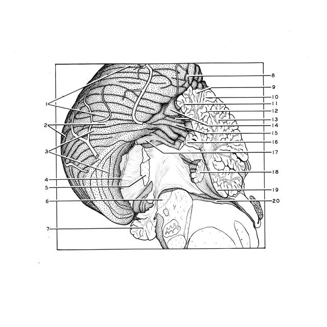
The inferior half of the pyramis has been cut away and its remaining part reflected downward. The prepyramidal fissure is thus revealed. The folia of the upper aspect of the pyramis (15) can be traced laterally into the biventral lobule (3). The lobulus gracilis (2) extends medially deep within the prepyramidal fissure to become continuous with the deepest folia of the tuber on the superior wall of the fissure. Folia of the inferior semilunar lobule (1) all converge medially to become continuous with the remaining folia of the tuber (11).
- Inferior semilunar lobe (crus II ansiform lobule)
- Gracile lobule (paramedian lobule)
- Biventral lobule (dorsal paraflocculus)
- Medullary center of lateral part of cerebellar tonsil
- Medullary center of medial part of cerebellar tonsil
- Restiform body (inferior cerebellar peduncle)
- Flocculus (dissected)
- Horizontal cerebellar sulcus
- Folium vermis
- Declive of cerebellum
- Tuber vermis
- Primary fissure
- Medullary substance of Culmen of monticulus
- Folia connecting inferior semilunar lobule and tuber vermis
- Folium connecting gracile lobule and tuber vermis
- Pyramid (partially removed and reflected)
- Folium connecting lateral part of tonsil with pyramid
- Nodulus
- Cerebellar lingula
- Median eminence rhomboid fossa