Exploration of the cerebellum and brain stem from above and behind
Rhomboid fossa of fourth ventricle
Stanford holds the copyright to the David L. Bassett anatomical images and has assigned
Creative Commons license Attribution-Share
Alike 4.0 International to all of the images.
For additional information regarding use and permissions,
please contact the Medical History Center.
Image #24-5
Exploration of the cerebellum and brain stem from above and behind
Rhomboid fossa of fourth ventricle
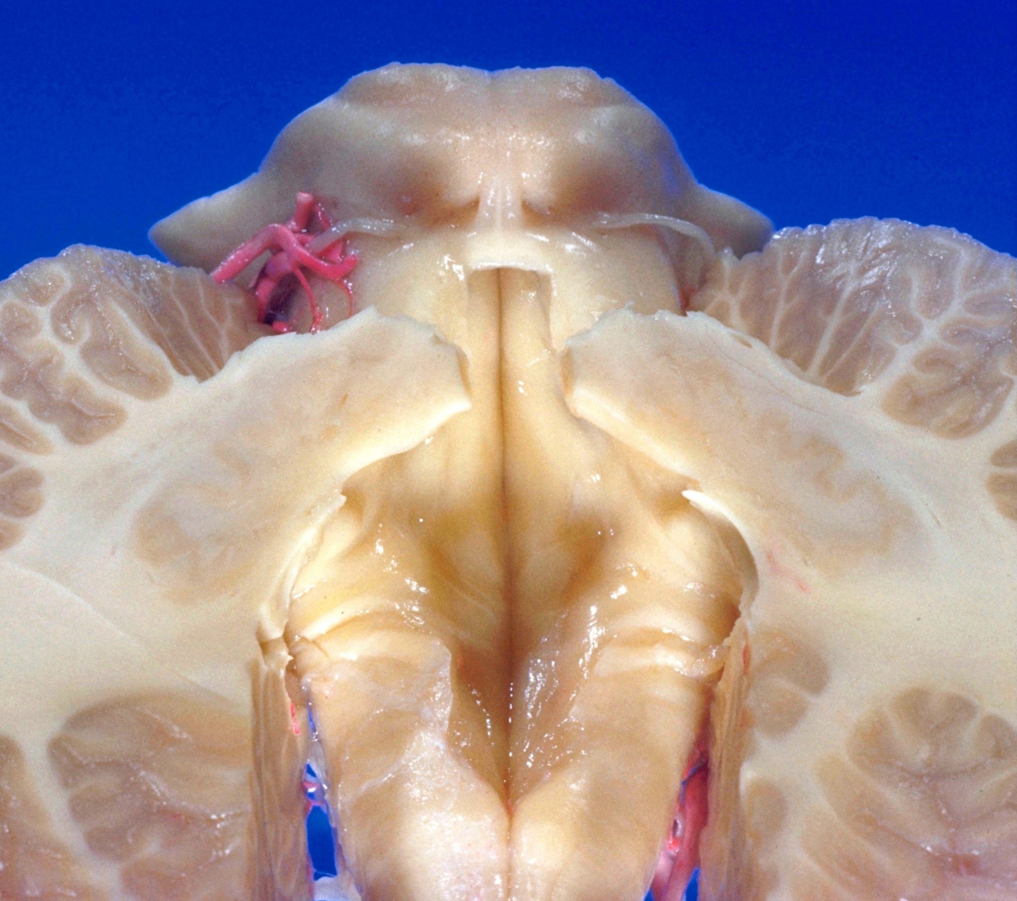
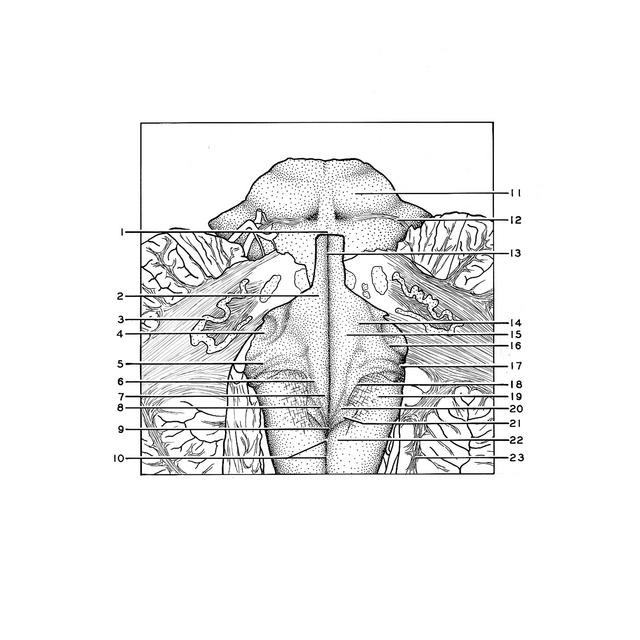
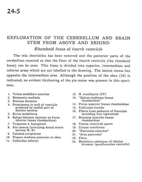
The tela chorioidea has been removed and the posterior parts of the cerebellum resected so that the floor of the fourth ventricle (the rhomboid fossa) can be seen. This fossa is divided into superior, intermediate and inferior areas which are not labelled in the drawing. The lateral recess lies opposite the intermediate area. Although the position of the obex (10) is indicated, no evident thickening of the pia mater was present in this specimen.
- Anterior medullary velum
- Median eminence
- Dentate nucleus
- Prominence in wall of ventricle produced by medial part of dentate nucleus
- Medullary striae
- Sulcus limitans (pointer on inferior fovea rhomboid fossae)
- Hypoglossal trigone
- Wing of gray matter (overlying dorsal motor nucleus nerve X)
- Calamus scriptorius (dorsal median sulcus)
- Posterior median fissure and obex
- Inferior colliculus
- Trochlear nerve (IV)
- Median sulcus of rhomboid fossa
- Superior fovea rhomboid fossa
- Facial colliculus
- Fibers from peduncle of flocculus ascending into tegmentum
- Lateral recess of rhomboid fossa
- Taenia fourth ventricle
- Restiform body (inferior cerebellar peduncle)
- "Funiculus separans"
- "Area postrema"
- Clava
- Medullary substance of biventral lobule (ventral paraflocculus)