Exploration of the cerebellum and brain stem from above and behind
Lingula and anterior medullary velum; tela chorioidea of fourth ventricle
Stanford holds the copyright to the David L. Bassett anatomical images and has assigned
Creative Commons license Attribution-Share
Alike 4.0 International to all of the images.
For additional information regarding use and permissions,
please contact the Medical History Center.
Image #24-3
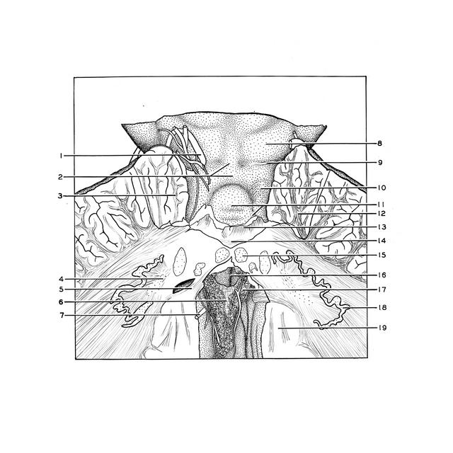
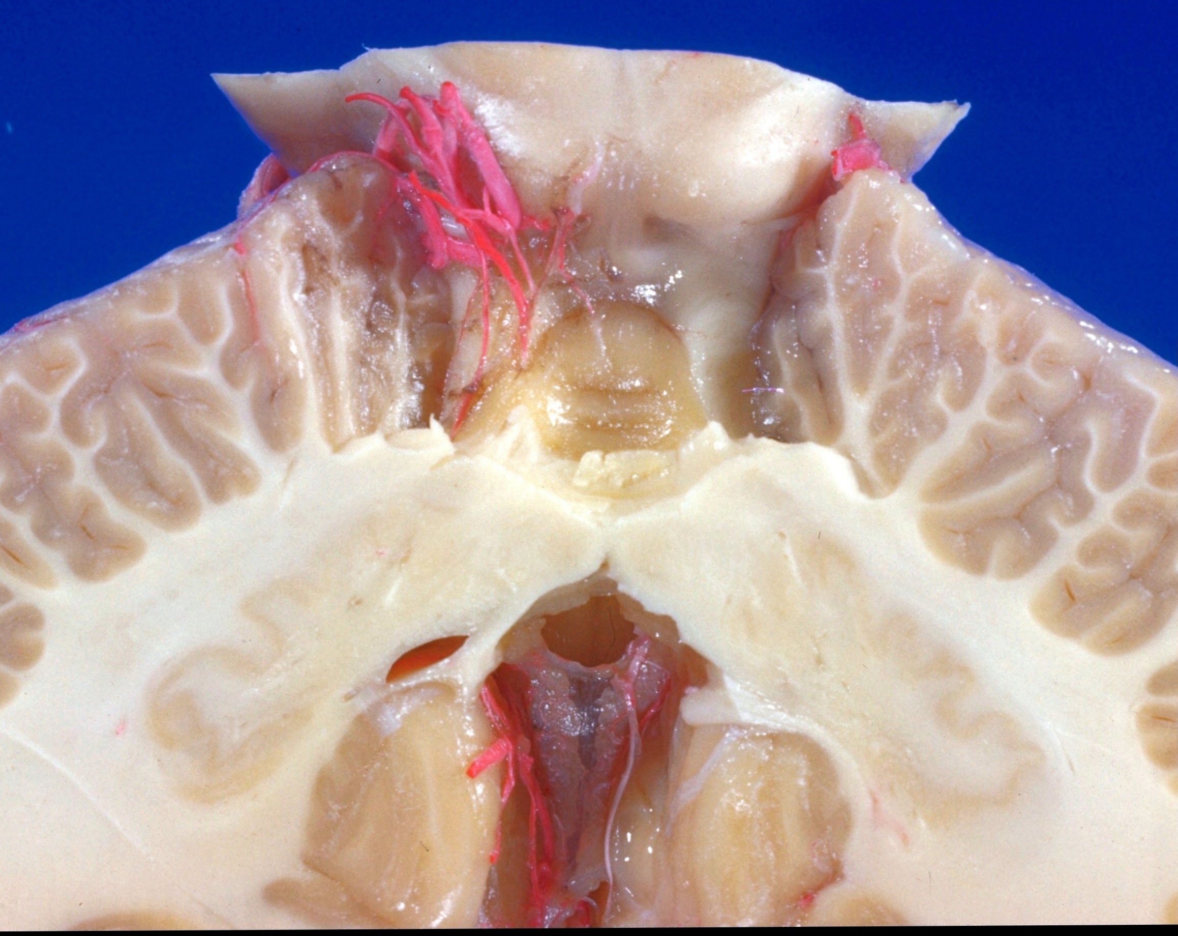
Exploration of the cerebellum and brain stem from above and behind
Lingula and anterior medullary velum; tela chorioidea of fourth ventricle
The lobulus centralis has been cut away to display the lingula (11). The terminal portion of the ventral spinocerebellar tract is visible on the right side in its ascending course across the brachium conjunctivum. Note the pigmentation of the meninges which have been left intact over the left brachium conjunctivum. Posteriorly the uvula and nodulus of the vermis have been removed. Below these the roof of the fourth ventricle is thin (tela chorioidea) and contains two folds of choroid plexus. The cavity of the ventricle (16) is seen through a tear in this membrane.
- Superior cerebellar artery
- Anterior medullary velum and frenulum veli
- Ala of central lobule (cut across)
- Hilus dentate nucleus
- Posterior medullary velum
- Tela chorioidea fourth ventricle
- Posterior inferior cerebellar artery
- Inferior colliculus
- Trochlear nerve (IV)
- Ventral spinocerebellar tract
- Lingula and cerebellar band of lingula
- Medullary center of anterior part quadrangular lobule (anterior semilunar lobule)
- Medullary center of central lobule (cut across)
- Commissural fibers
- Fastigial nucleus
- Fourth ventricle (visible through tear in tela chorioidea)
- Choroid plexus (branches of the choroidal artery appear white instead of red here)
- Dentate nucleus
- Tonsil (ventral paraflocculus)