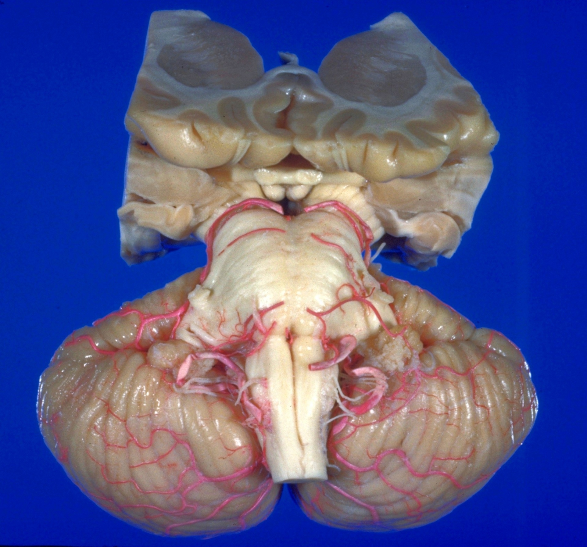
The Surfaces of the cerebellum and brain stem
Inferior aspect
Stanford holds the copyright to the David L. Bassett anatomical images and has assigned
Creative Commons license Attribution-Share
Alike 4.0 International to all of the images.
For additional information regarding use and permissions,
please contact the Medical History Center.
Image #23-5
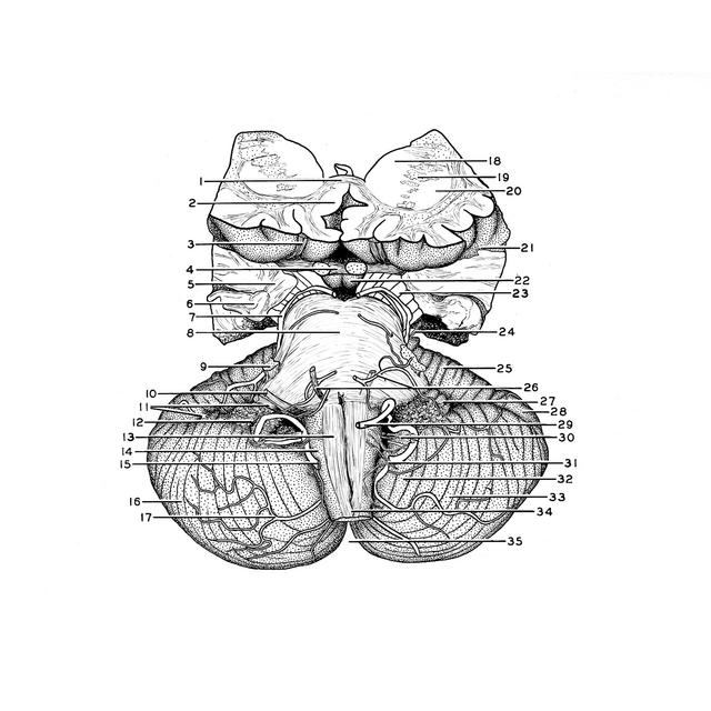
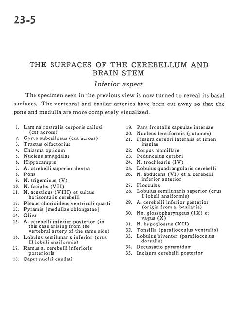
The Surfaces of the cerebellum and brain stem
Inferior aspect
The specimen seen in the previous view is now turned to reveal its basal surfaces. The vertebral and basilar arteries have been cut away so that the pons and medulla are more completely visualized.
- Rostral lamina of corpus callosum (cut across)
- Subcallosal gyrus (cut across)
- Olfactory tract
- Optic chiasm
- Amygdaloid nucleus
- Hippocampus
- Superior cerebellar artery right
- Pons
- Trigeminal nerve (V)
- Facial nerve (VII)
- Vestibulocochlear nerve (VIII) and horizontal cerebellar sulcus
- Choroid plexus fourth ventricle
- Pyramid [medullae oblongatae]
- Olive
- Posterior inferior cerebellar artery (in this case arising from the vertebral artery of the same side)
- Inferior semilunar lobe (crus II ansiform lobule)
- Branch posterior inferior cerebellar artery (PICA)
- Head of caudate nucleus
- Frontal part internal capsule
- Lentiform nucleus (putamen)
- Lateral cerebral fissure and limen insulae
- Mamillary body
- Cerebral peduncle
- Trochlear nerve (IV)
- Quadrangular lobe of cerebellum
- Abducens nerve (Vl) and anterior inferior cerebellar artery
- Flocculus
- Superior semilunar lobe (crus I ansiform lobule)
- Posterior inferior cerebellar artery (origin from basilar artery)
- Glossopharyngeal (IX) and vagus (X) nerves
- Hypoglossal nerve (XII)
- Tonsil (ventral paraflocculus)
- Biventral lobule (dorsal paraflocculus)
- Pyramidal decussation
- Posterior cerebellar incisura