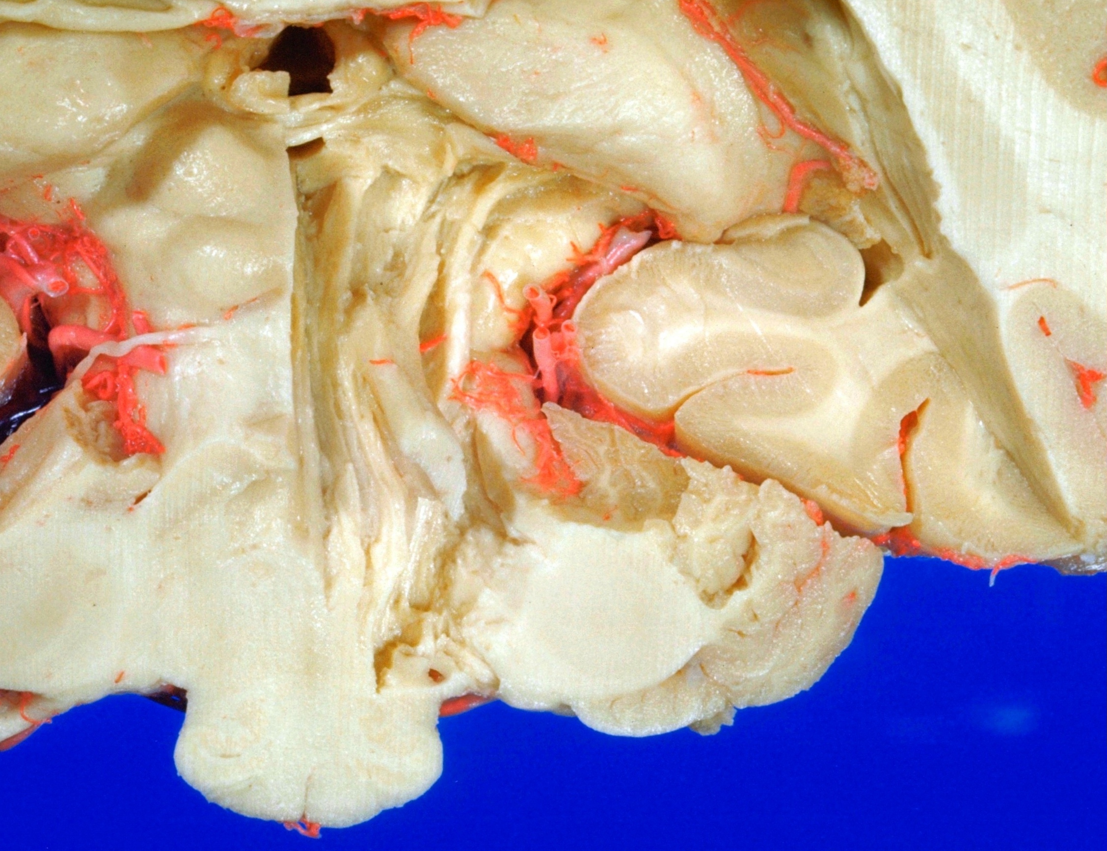
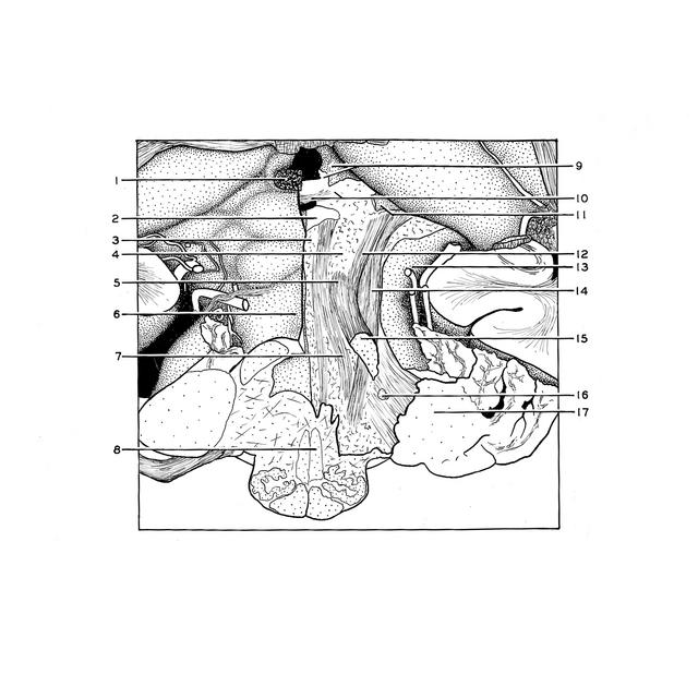
Exploration of a brain cut in frontal section at the splenium of the corpus callosum
Central tegmental tract; posterior commissure; termination of medial lemniscus within thalamus
Stanford holds the copyright to the David L. Bassett anatomical images and has assigned
Creative Commons license Attribution-Share
Alike 4.0 International to all of the images.
For additional information regarding use and permissions,
please contact the Medical History Center.
Image #22-6
Exploration of a brain cut in frontal section at the splenium of the corpus callosum
Central tegmental tract; posterior commissure; termination of medial lemniscus within thalamus
The posterior commissure (10) has been exposed by removing the remainder of the right superior colliculus. Fibers from this commissure are faintly visible as they course into the pretectal region immediately lateral to the aqueduct. The medial lemniscus can be traced into the ventral posterior lateral thalamic nucleus. In the tegmental part of the pons and mesencephalon a large mass of longitudinally coursing fibers, the central tegmental tract, is uncovered. The more medial of these fibers lie in a position corresponding to that of the medial longitudinal fasciculus but they cannot be clearly separated from the remainder.
- Pineal body (cut off)
- Central gray matter
- Oculomotor nucleus (III)
- Tissue overlying red nucleus
- Level at which brachium conjunctivum (superior cerebellar peduncle) decussates
- Anterior medullary velum
- Central tegmental tract
- Position of medial lemniscus (cut across)
- Habenula and habenular commissure (cut across)
- Posterior commissure and pretectal region
- Brachium of superior colliculus (cut across)
- Uncrossed fibers of brachium conjunctivum (superior cerebellar peduncle)
- Cerebral peduncle
- Medial lemniscus
- Brachium conjunctivum (superior cerebellar peduncle) (cut across)
- Fibers of trigeminal nerve (cut across)
- Brachium pontis (middle cerebellar peduncle)