Exploration of a brain cut in frontal section at the splenium of the corpus callosum
Cerebral aqueduct, tegmentum of mesencephalon and medial lemniscus
Stanford holds the copyright to the David L. Bassett anatomical images and has assigned
Creative Commons license Attribution-Share
Alike 4.0 International to all of the images.
For additional information regarding use and permissions,
please contact the Medical History Center.
Image #22-5
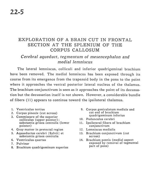
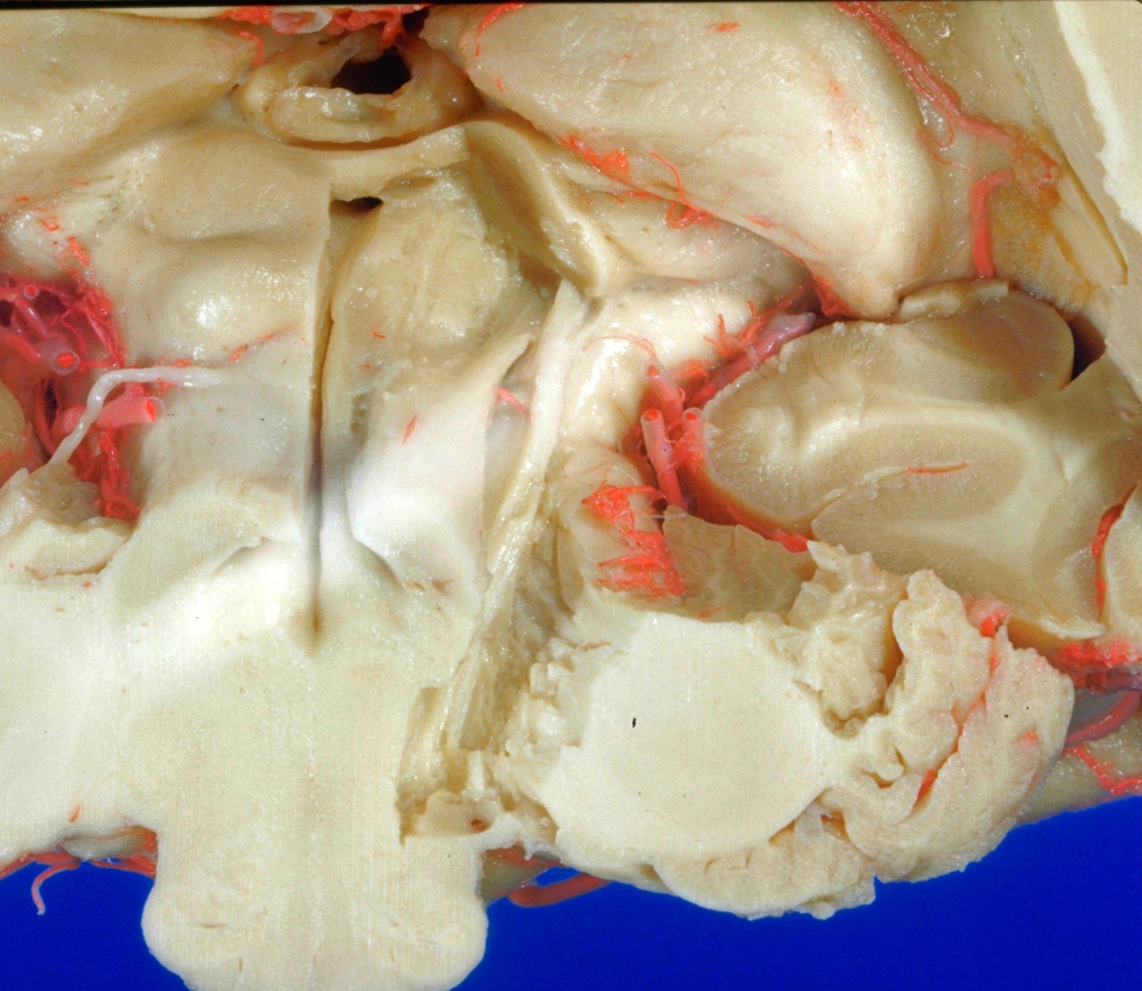
Exploration of a brain cut in frontal section at the splenium of the corpus callosum
Cerebral aqueduct, tegmentum of mesencephalon and medial lemniscus
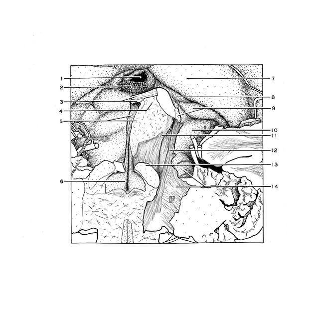
The lateral lemniscus, colliculi and inferior quadrigeminal brachium have been removed. The medial lemniscus has been exposed through its course from its emergence from the trapezoid body in the pons to the point where it approaches the ventral posterior lateral nucleus of the thalamus. The brachium conjunctivum is seen as it approaches the point of its decussation but the decussation itself is not shown. However, a considerable bundle of fibers (11) appears to continue toward the ipsilateral thalamus.
- Third ventricle
- Pineal body (cut across)
- Commissure of the superior colliculus (upper pointer) Central gray matter (lower pointer)
- Gray matter in pretectal region
- Cerebral aqueduct and central gray matter
- Fourth ventricle
- Pulvinar
- Brachium of superior colliculus
- Medial geniculate body and cut end of brachiurn of inferior colliculus
- Cerebral peduncle
- Ipsilateral fibers of brachium conjunctivum (superior cerebellar peduncle)
- Medial lemniscus
- Brachium conjunctivum (superior cerebellar peduncle) (cut across)
- Brachium pontis (middle cerebellar peduncle) (medial aspect exposed by removal of tegmental part of pons)