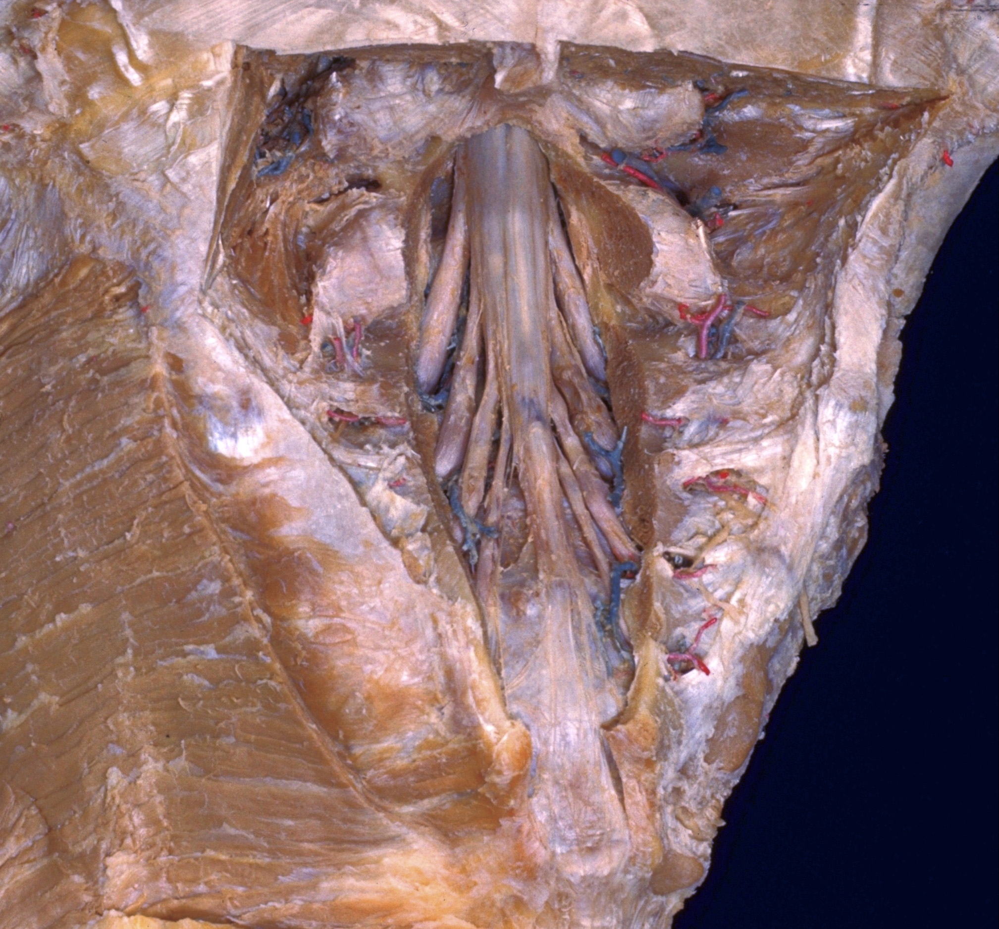
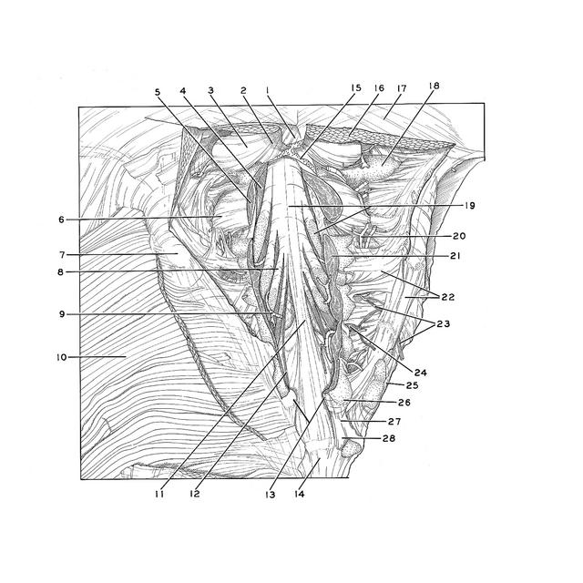
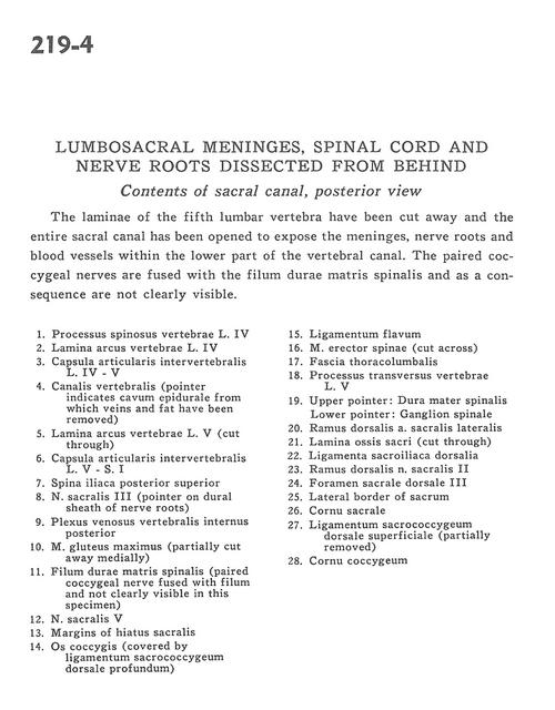
Lumbosacral meninges, spinal cord and nerve roots dissected from behind
Contents of sacral canal, posterior view
Stanford holds the copyright to the David L. Bassett anatomical images and has assigned
Creative Commons license Attribution-Share
Alike 4.0 International to all of the images.
For additional information regarding use and permissions,
please contact the Medical History Center.
Image #219-4
Lumbosacral meninges, spinal cord and nerve roots dissected from behind
Contents of sacral canal, posterior view
The laminae of the fifth lumbar vertebra have been cut away and the entire sacral canal has been opened to expose the meninges, nerve roots and blood vessels within the lower part of the vertebral canal. The paired coccygeal nerves are fused with the filum durae matris spinalis and as a consequence are not clearly visible.
- Spinous process vertebra L. IV
- Lamina (arch of vertebra) L. IV
- Intervertebral joint capsule L. IV - V
- Vertebral canal (pointer indicates epidural space from which veins and fat have been removed)
- Lamina (arch of vertebra) L. V (cut through)
- Intervertebral joint capsule L. V - S. I
- Posterior superior iliac spine
- Sacral nerve III (pointer on dural sheath of nerve roots)
- Posterior internal vertebral venous plexus
- Gluteus maximus muscle (partially cut away medially)
- Dural filum terminale (paired coccygeal nerve fused with filum and not clearly visible in this specimen)
- Sacral nerve V
- Margins of sacral hiatus
- Coccyx (covered by deep dorsal sacrococcygeal ligament)
- Ligamentum flavum
- Erector spinae muscle (cut across)
- Thoracolumbar fascia
- Transverse process of vertebra L. V
- Upper pointer: Dura mater Lower pointer: Spinal ganglion
- Dorsal branch lateral sacral artery
- Lamina sacrum (cut through)
- Dorsal sacroiliac ligament
- Dorsal branch sacral nerve II
- Posterior sacral foramen III
- Lateral border of sacrum
- Sacral cornu (horn)
- Dorsal superficial sacrococcygeal ligament (partially removed)
- Coccygeal cornu (horn)