Thoracic meninges, spinal cord and nerve roots dissected in relation to vertebral column
Intrathoracic structures related to vertebral column, posterior view
Stanford holds the copyright to the David L. Bassett anatomical images and has assigned
Creative Commons license Attribution-Share
Alike 4.0 International to all of the images.
For additional information regarding use and permissions,
please contact the Medical History Center.
Image #219-3
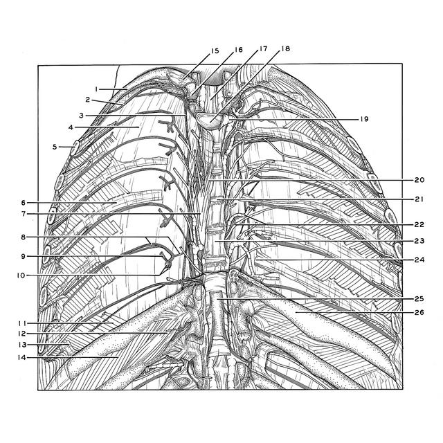
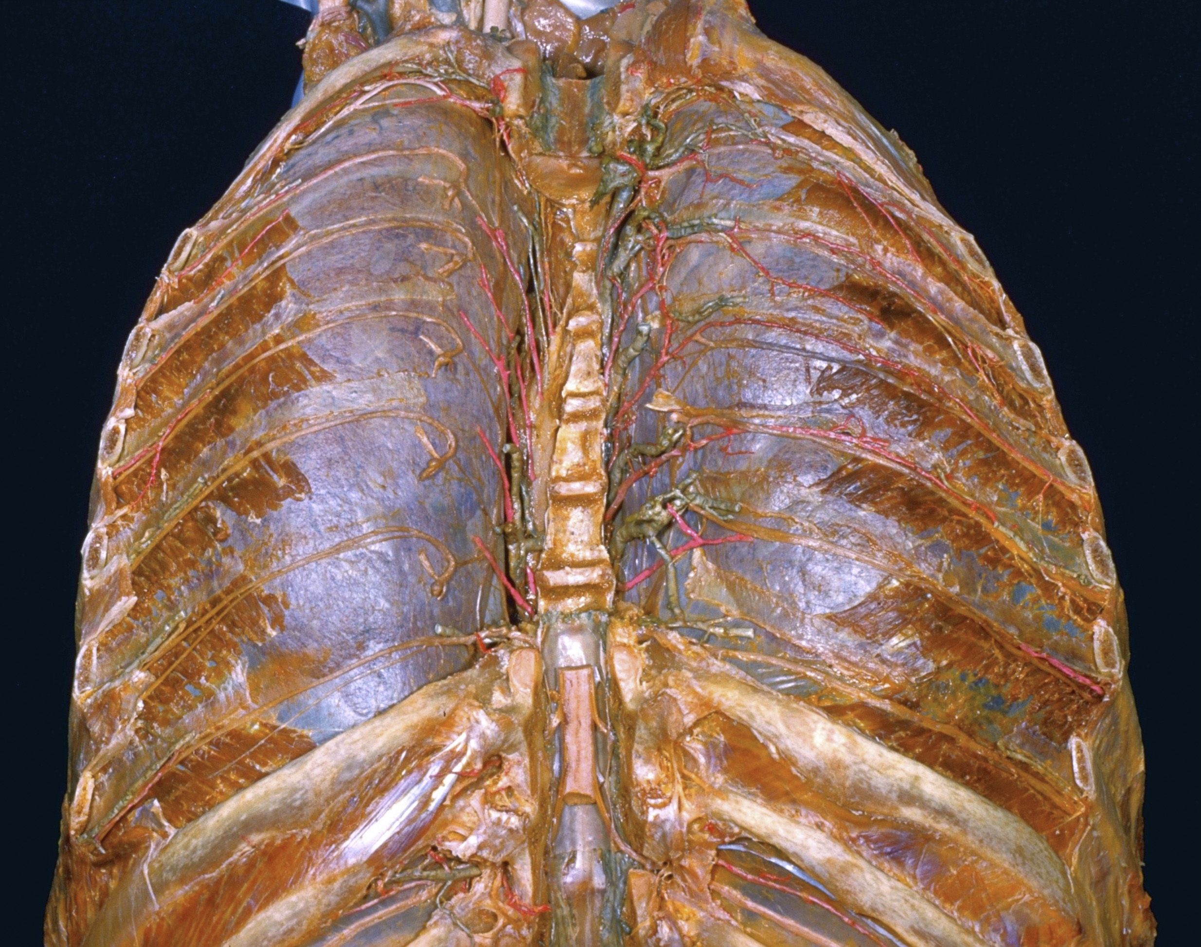
Thoracic meninges, spinal cord and nerve roots dissected in relation to vertebral column
Intrathoracic structures related to vertebral column, posterior view
Ribs and vertebral bodies have been resected bilaterally between the second and ninth thoracic levels. The periosteum (6) which covered the inner surfaces of the ribs has been preserved in most areas. The anterior longitudinal ligament (23), with remnants of the intervertebral discs attached,has also been retained in part. The lungs have been inflated and are visible through the intact costal pleura. The proximal parts of the III-VII thoracic nerves have been positioned on the pleura in such a way that their dorsal and ventral roots, dorsal rami and communications with the sympathetic trunk are visible. These components are labeled for the left seventh thoracic nerve (8,9,10). The intercostal arteries and veins have been cut off in various ways.
- Rib II
- Intercostal nerve II
- Sympathetic trunk
- Costal pleura
- Rib III (cut off)
- Periosteum of sixth rib
- Thoracic aorta
- Left pointer: Intercostal. nerve VII Right pointer: Ramus communicans
- Dorsal branch thoracic nerve VII
- Left pointer: Spinal ganglion and dorsal roots Right pointer: Ventral root
- Innermost intercostal muscle
- Levator costarum brevis muscle
- Internal intercostal membrane
- External intercostal muscle
- Transverse process of vertebra Th. II
- Inferior articular process
- Posterior longitudinal ligament
- Intervertebral disc Th. II - III
- Veins of third intervertebral foramen (bones removed)
- Vertebromediastinal suspensory ligament
- Anulus fibrosus intervertebral disc Th. V - VI
- Posterior intercostal artery and vein VI
- Anterior longitudinal ligament
- Radiate ligament (preserved with periosteum of rib)
- Spinal cord
- Rib IX