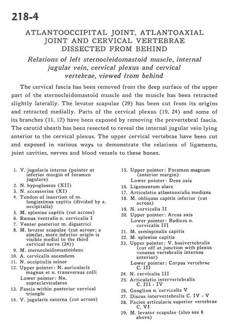
Atlantooccipital joint, atlantoaxial joint and cervical vertebrae dissected from behind
Relations of left sternocleidomastoid muscle, internal jugular vein, cervical plexus and cervical vertebrae, viewed from behind
Stanford holds the copyright to the David L. Bassett anatomical images and has assigned
Creative Commons license Attribution-Share
Alike 4.0 International to all of the images.
For additional information regarding use and permissions,
please contact the Medical History Center.
Image #218-4
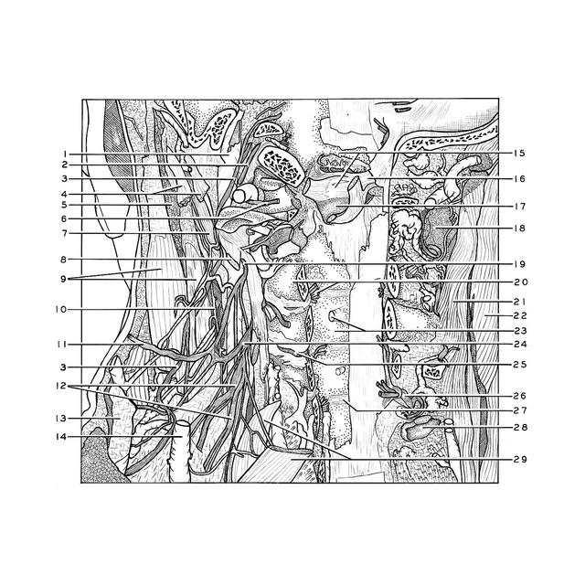
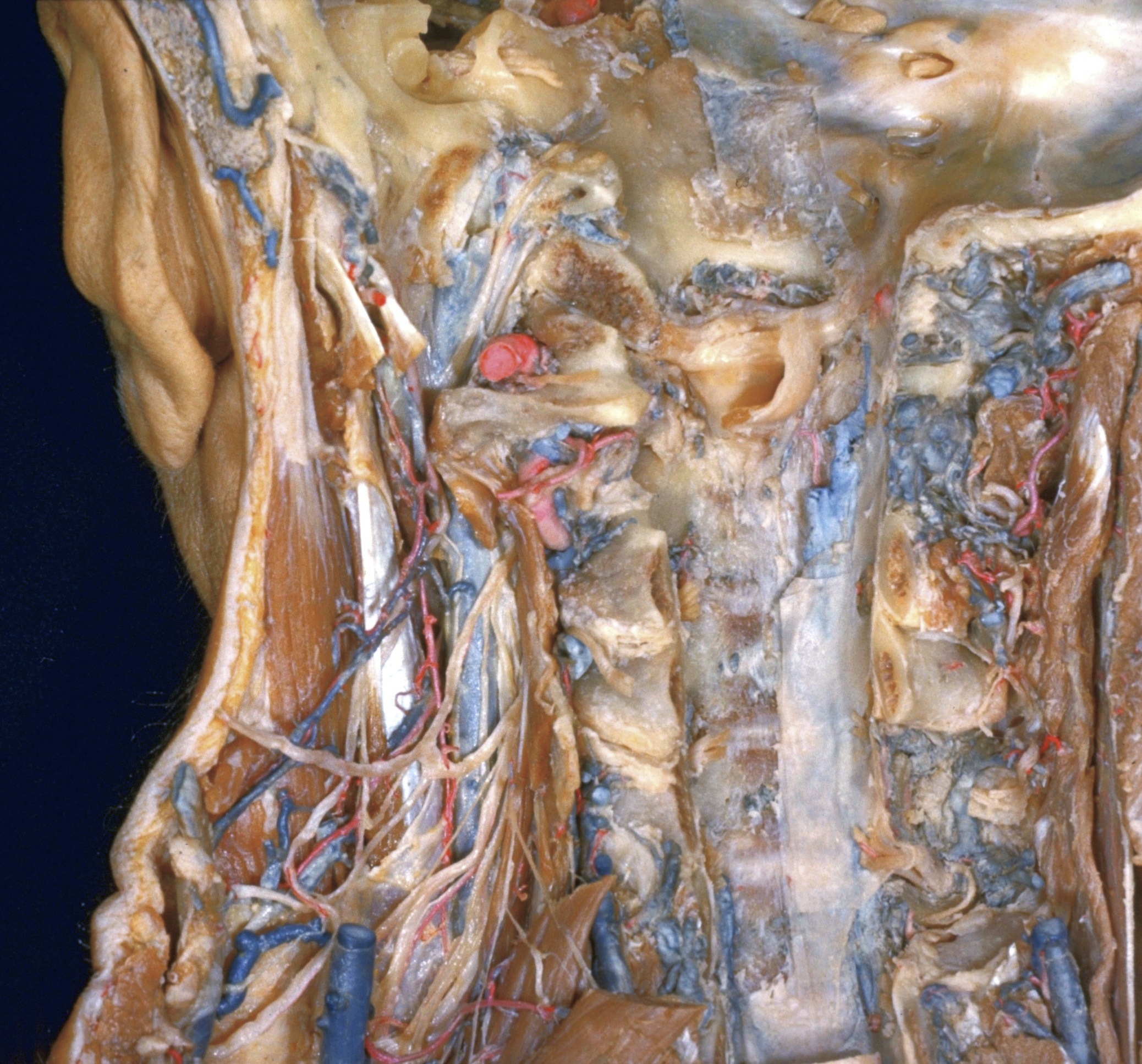
Atlantooccipital joint, atlantoaxial joint and cervical vertebrae dissected from behind
Relations of left sternocleidomastoid muscle, internal jugular vein, cervical plexus and cervical vertebrae, viewed from behind
The cervical fascia has been removed from the deep surface of the upper part of the sternocleidomastoid muscle and the muscle has been retracted slightly laterally. The levator scapulae (29) has been cut from its origins and retracted medially. Parts of the cervical plexus (19,24) and some of its branches (11,12) have been exposed by removing the prevertebral fascia. The carotid sheath has been resected to reveal the internal jugular vein lying anterior to the cervical plexus. The upper cervical vertebrae have been cut and exposed in various ways to demonstrate the relations of ligaments, joint cavities, nerves and blood vessels to these bones.
- Internal jugular vein (pointer at inferior margin of jugular foramen)
- Hypoglossal nerve (XII)
- Accessory nerve (XI)
- Tendon of insertion of longissimus capitis muscle (divided by occipital artery)
- Splenius capitis muscle (cut across)
- Ventral branch cervical nerve I
- Posterior belly of digastric muscle
- Levator scapulae muscle (cut across a similar, more inferior origin is visible medial to the third cervical nerve (24))
- Sternocleidomastoid muscle
- Ascending cervical artery
- Lesser occipital nerve
- Upper pointer: Greater auricular nerve and superficial transverse nerve Lower pointer: Supraclavicular nerves
- Fascia within posterior cervical triangle
- External jugular vein (cut across)
- Upper pointer: Foramen magnum (anterior margin) Lower pointer: Dens (axis)
- Alar ligament
- Median atlantoaxial joint
- Obliquus capitis inferior muscle (cut across)
- Cervical nerve II
- Upper pointer: Arch of axis Lower pointer: Roots of cervical nerve III
- Semispinalis capitis muscle
- Splenius capitis muscle
- Upper pointer: Basivertebral vein (cut off at junction with anterior internal vertebral venous plexus) Lower pointer: Body of vertebra C. III
- Cervical nerve III
- Intervertebral joint nerve III-IV
- Ganglion cervical nerve V
- Intervertebral disc C. IV - V
- Superior articular surface vertebra C. VI
- Levator scapulae muscle (also see 8 above)