Atlantooccipital joint, atlantoaxial joint and cervical vertebrae dissected from behind
Tectorial membrane; atlantooccipital joint
Stanford holds the copyright to the David L. Bassett anatomical images and has assigned
Creative Commons license Attribution-Share
Alike 4.0 International to all of the images.
For additional information regarding use and permissions,
please contact the Medical History Center.
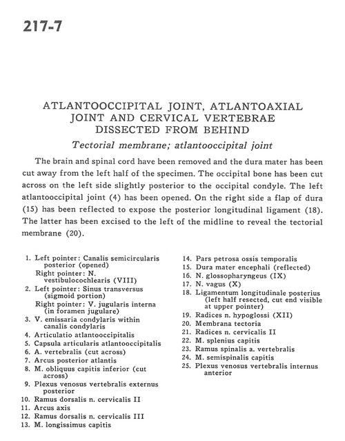
Image #217-7
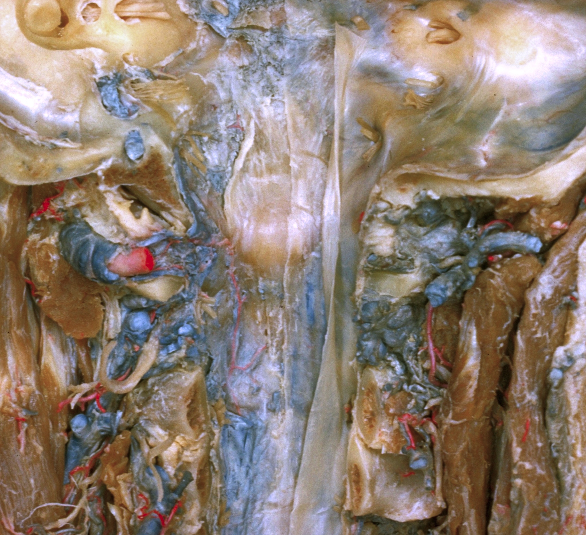
Atlantooccipital joint, atlantoaxial joint and cervical vertebrae dissected from behind
Tectorial membrane; atlantooccipital joint
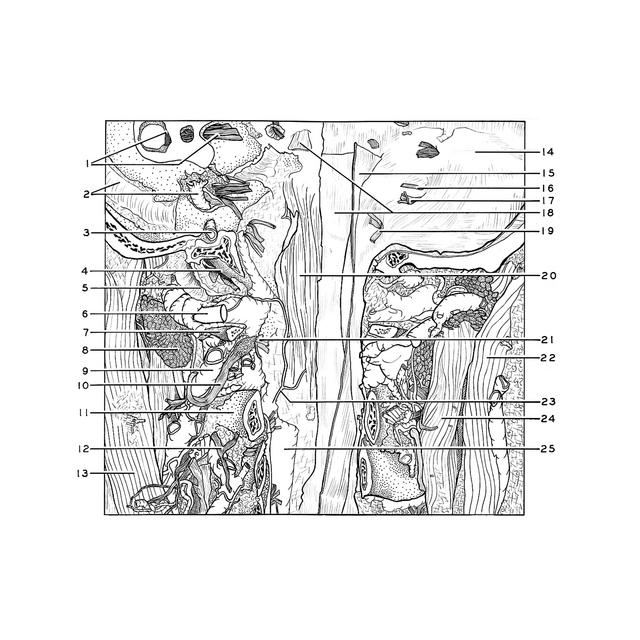
The brain and spinal cord have been removed and the dura mater has been cut away from the left half of the specimen. The occipital bone had been cut across on the left side slightly posterior to the occipital condyle. The left atlantooccipital joint (4) has been opened. On the right side a flap of dura (15) has been reflected to expose the posterior longitudinal ligament (18). The latter has been excised to the left of the midline to reveal the tectorial membrane (20).
- Left pointer: Posterior semicircular canal (opened) Right pointer: Vestibulocochlear nerve (VIII)
- Left pointer: Transverse sinus (sigmoid portion) Right pointer: Internal jugular vein (in jugular foramen)
- Condylar emissary vein within condylar canal
- Atlanto-occipital joint
- Atlanto-occipital joint capsule
- Vertebral artery (cut across)
- Posterior arch of atlas
- Obliquus capitis inferior muscle (cut across)
- Posterior external vertebral venous plexus
- Dorsal branch cervical nerve II
- Arch of axis
- Dorsal branch cervical nerve III
- Longissimus capitis muscle
- Petrosal part temporal bone
- Dura mater (reflected)
- Glossopharyngeal nerve (IX)
- Vagus nerve (X)
- Posterior longitudinal ligament (left half resected, cut end visible at upper pointer)
- Roots of hypoglossal nerve (XII)
- Tectorial membrane
- Roots of cervical nerve II
- Splenius capitis muscle
- Spinal branch vertebral artery
- Semispinalis capitis muscle
- Anterior internal vertebral venous plexus