Dissection of thoracic and lumbosacral regions of back from a posterior approach
Superficial structures and external layer of muscles of back, general view
Stanford holds the copyright to the David L. Bassett anatomical images and has assigned
Creative Commons license Attribution-Share
Alike 4.0 International to all of the images.
For additional information regarding use and permissions,
please contact the Medical History Center.
Image #212-1
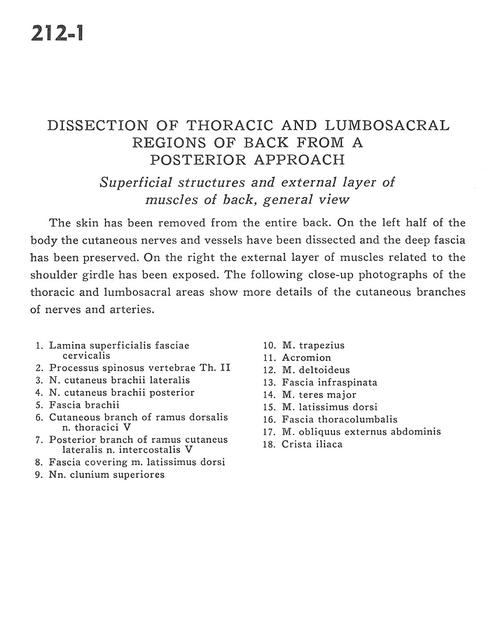
Dissection of thoracic and lumbosacral regions of back from a posterior approach
Superficial structures and external layer of muscles of back, general view
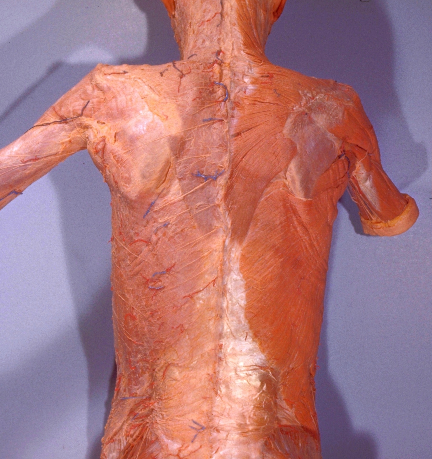
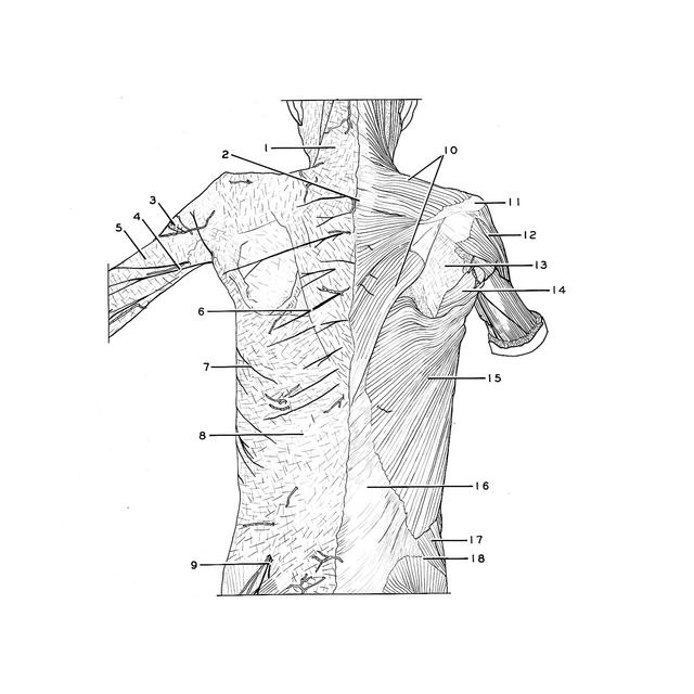
The skin has been removed form the entire back. On the left half of the body the cutaneous nerves and vessels have been dissected and the deep fascia has been preserved. On the right the external layer of muscles related to the shoulder girdle has been exposed. The following close-up photographs of the thoracic and lumbosacral areas show more details of the cutaneous branches of nerves and arteries.
- Superficial fascia cervical surface
- Spinous process vertebra Th. II
- Lateral brachial cutaneous nerve
- Posterior brachial cutaneous nerve
- Brachial fascia
- Dorsal cutaneous branch thoracic nerve V
- Posterior branch of lateral cutaneous branch intercostal nerve V
- Fascia covering latissimus dorsi muscle
- Superior cluneal nerves
- Trapezius muscle
- Acromion
- Deltoid muscle
- Fascia infraspinata
- Teres major muscle
- Latissimus dorsi muscle
- Thoracolumbar fascia
- External oblique muscle
- Iliac crest