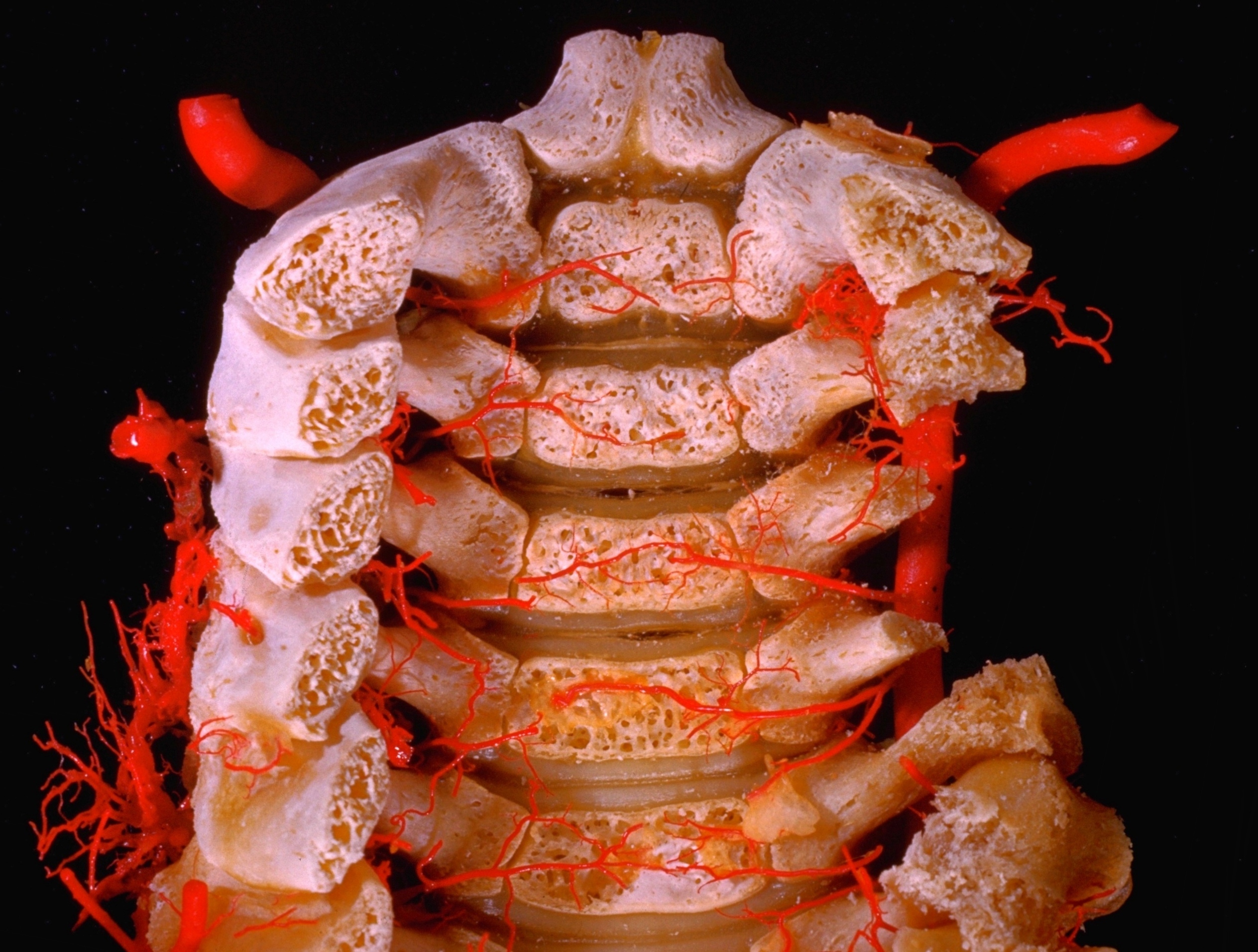
Arteries of vertebral column of one
Year-old infant - Arteries within cervical part of vertebral canal, posterior view
Stanford holds the copyright to the David L. Bassett anatomical images and has assigned
Creative Commons license Attribution-Share
Alike 4.0 International to all of the images.
For additional information regarding use and permissions,
please contact the Medical History Center.
Image #211-3
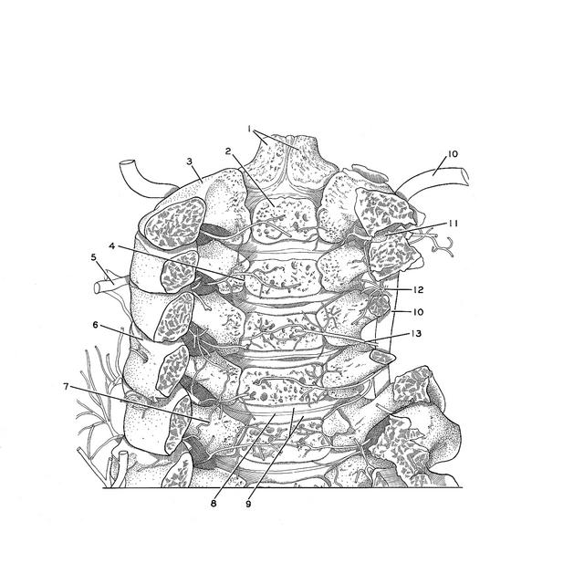
Arteries of vertebral column of one
Year-old infant - Arteries within cervical part of vertebral canal, posterior view
The vertebral arches have been trimmed away to reveal the vertebral canal in the cervical region. On the right side of the specimen the pedicles of the fourth and fifth vertebrae have been cut across so that the right vertebral artery (10) is visible with three of its segmental spinal branches. The atlas has been entirely removed from the specimen.
- Dens (axis) (two ossification centers visible)
- Ossification center for body of axis
- Arch of axis
- Area of fusion of ossification centers for body and neural arch of third cervical vertebra
- External vertebral venous plexus (injected)
- Superior articular surface vertebra C. V
- Spinal branch vertebral artery (radicular branch which followed roots of seventh cervical nerve)
- Intervertebral disc C. V - VI
- Unossified cartilage of bodies of vertebra C. V and C. VI
- Vertebral artery
- Superior articular process vertebra C. III
- Spinal branch vertebral artery (dorsal branch to vertebral arch)
- Spinal branch vertebral artery (ventral branch to vertebral body)