Radiography
Radiograph of head and neck, left oblique view
Stanford holds the copyright to the David L. Bassett anatomical images and has assigned
Creative Commons license Attribution-Share
Alike 4.0 International to all of the images.
For additional information regarding use and permissions,
please contact the Medical History Center.
Image #210-1
Radiography
Radiograph of head and neck, left oblique view
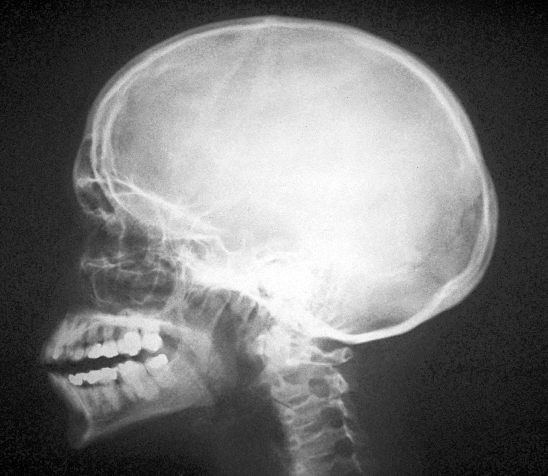
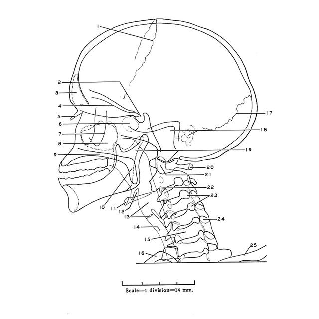
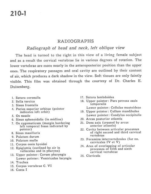
The head is turned to the right in this view of a living female subject and as a result the cervical vertebrae lie in various degrees of rotation. The lower vertebrae are more nearly in the anteroposterior position than the upper ones. The respiratory passages and the oral cavity are outlined by their content of air, which produces a dark shadow in the view. Soft tissues are only faintly visible. This film was obtained through the courtesy of Dr. Charles E. Duisenberg.
- Coronal suture
- Sella turcica
- Frontal sinus
- Superior wall of orbit (pointer indicates left orbit)
- Nasal bone
- Sphenoidal sinus (in midline)
- Zygomatic bone (margin bordering left temporal fossa indicated by pointer)
- Maxillary sinus
- Hard palate
- Soft palate
- Body hyoid bone
- Epiglottis (outlined by air in valleculae and in pharynx)
- Upper pointer: Pharyngeal cavity Lower pointer: Ventricle of larynx
- Trachea
- Body of vertebra C. VI
- Rib I
- Lambdoid suture
- Upper pointer: Petrosal part temporal bone Lower pointer: Mastoid cell
- Upper pointer: Neck of mandible Lower pointer: Occipital condyle
- Posterior arch of atlas
- Dens (axis) (crossed by anterior arch of atlas)
- Cavity between articular processes of right second and third cervical vertebrae
- Intervertebral foramina (for cervical nerves lV and V)
- Area of overlapping of articular processes of fifth and sixth cervical vertebrae
- Clavicle