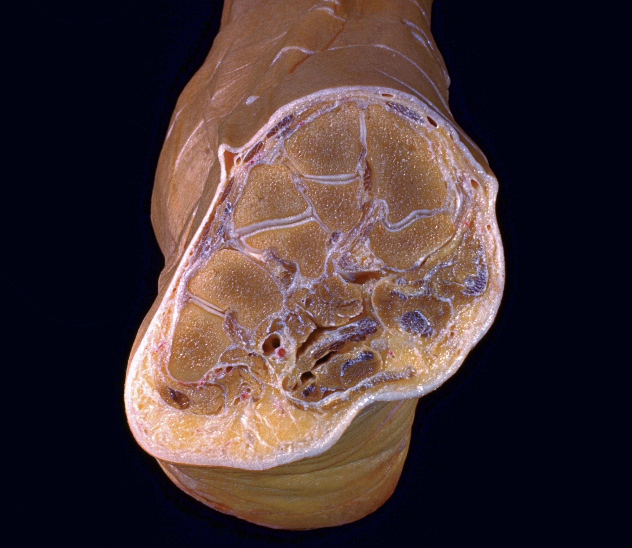
Cross sections of right foot
Section of foot through tarsometatarsal joints, viewed from in front
Stanford holds the copyright to the David L. Bassett anatomical images and has assigned
Creative Commons license Attribution-Share
Alike 4.0 International to all of the images.
For additional information regarding use and permissions,
please contact the Medical History Center.
Image #204-7
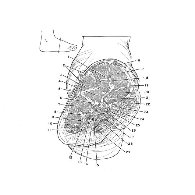
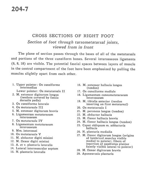
Cross sections of right foot
Section of foot through tarsometatarsal joints, viewed from in front
The plane of section passes through the bases of all of the metatarsals and portions of the three cuneiform bones. Several interosseous ligaments (6, 8, 18) are visible. The potential fascial spaces between layers of muscle in the central compartment of the foot have been emphasized by pulling the muscles slightly apart from each other.
- Upper pointer: Intermediate cuneiform bone Lower pointer: 1st metatarsal bone
- Extensor digitorum longus muscle (tendons covered by fascia of dorsalis pedis)
- Lateral cuneiform bone
- 2nd metatarsal bone
- Extensor digitorum brevis muscle
- Metatarsal interosseous ligament
- 5th metatarsal bone
- Metatarsal interosseous ligament
- Interosseous muscle
- 5th metatarsal bone
- Abductor digiti minimi muscle
- Flexor digiti minimi muscle
- Lateral plantar artery and vein
- Lateral intermuscular septum
- Lateral plantar nerve
- Extensor hallucis longus muscle (tendon)
- Medial cuneiform bone
- Cuneometatarsal interosseous ligament
- Tibialis anterior muscle (tendon inserting on 1st metatarsal)
- metatarsal bone
- Peroneus longus muscle (tendon)
- Abductor hallucis muscle
- Flexor hallucis brevis muscle
- Flexor hallucis longus muscle (tendon)
- Oblique head of adductor hallucis muscle
- Medial plantar nerve
- Flexor digitorum longus muscle (origins of lumbrical muscles visible medial to pointer fibers of insertion of quadratus plantae faintly visible lateral to pointer)
- Flexor digitorum brevis muscle
- Plantar aponeurosis