
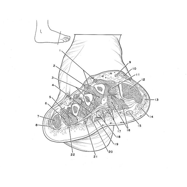
Cross sections of right foot
Section of foot through distal ends of metatarsals, viewed from in front
Stanford holds the copyright to the David L. Bassett anatomical images and has assigned
Creative Commons license Attribution-Share
Alike 4.0 International to all of the images.
For additional information regarding use and permissions,
please contact the Medical History Center.
Image #204-5
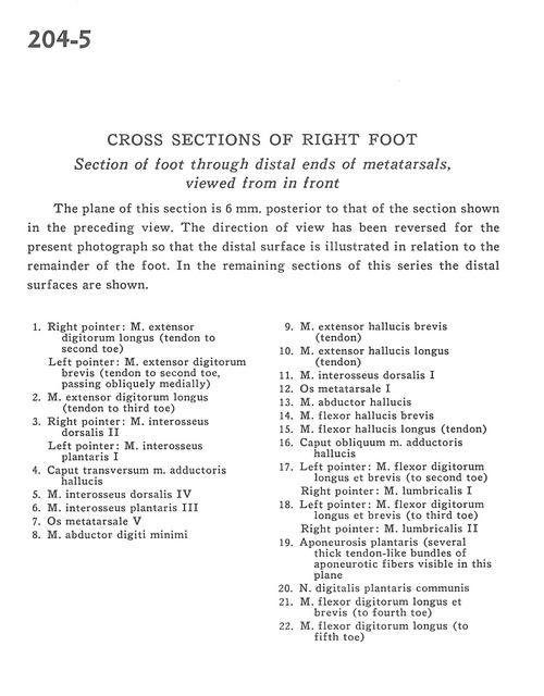
Cross sections of right foot
Section of foot through distal ends of metatarsals, viewed from in front
The plane of this section is 6 mm. posterior to that of the section shown in the preceding view. The direction of view has been reversed for the present photograph so that the distal surface is illustrated in relation to the remainder of the foot. In the remaining sections of this series the distal surfaces are shown.
- Right pointer: Extensor digitorum longus muscle (tendon to 2nd toe) Left pointer: Extensor digitorum brevis muscle (tendon to 2nd toe passing obliquely medially)
- Extensor digitorum longus muscle (tendon to 3rd toe)
- Right pointer: 2nd dorsal interosseous muscIe Left pointer: 1st plantar interosseous muscle
- Transverse head of adductor hallucis muscle
- 4th dorsal interosseus muscle
- 3rd plantar interosseous muscle
- 5th metatarsal bone
- Abductor digiti minimi muscle
- Extensor hallucis brevis muscle (tendon)
- Extensor hallucis longus muscle (tendon)
- 1st dorsal interosseus muscle
- Metatarsal bone
- Abductor hallucis muscle
- Flexor hallucis brevis muscle
- Flexor hallucis longus muscle (tendon)
- Oblique head of adductor hallucis muscle
- Left pointer: Flexor digitorum longus and brevis muscles (to 2nd toe) Right pointer: 1st lumbrical muscle
- Left pointer: Flexor digitorum longus and brevis muscles (to 3rd toe) Right pointer: 2nd lumbrical muscle
- Plantar aponeurosis (several thick tendon-like bundles of aponeurotic fibers visible in this plane)
- Common plantar digital nerve
- Flexor digitorum longus muscle et brevis (to 4th toe)
- Flexor digitorum longus muscle (to 5th toe)