Joints of left ankle and foot
Ligaments of plantar aspect of foot, deep layer
Stanford holds the copyright to the David L. Bassett anatomical images and has assigned
Creative Commons license Attribution-Share
Alike 4.0 International to all of the images.
For additional information regarding use and permissions,
please contact the Medical History Center.
Image #202-6
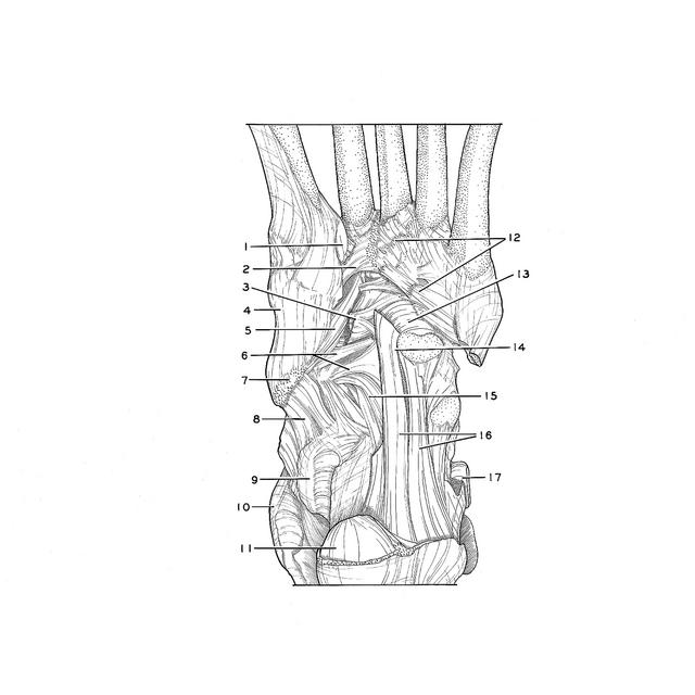
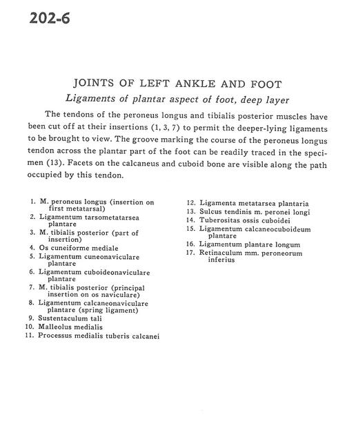
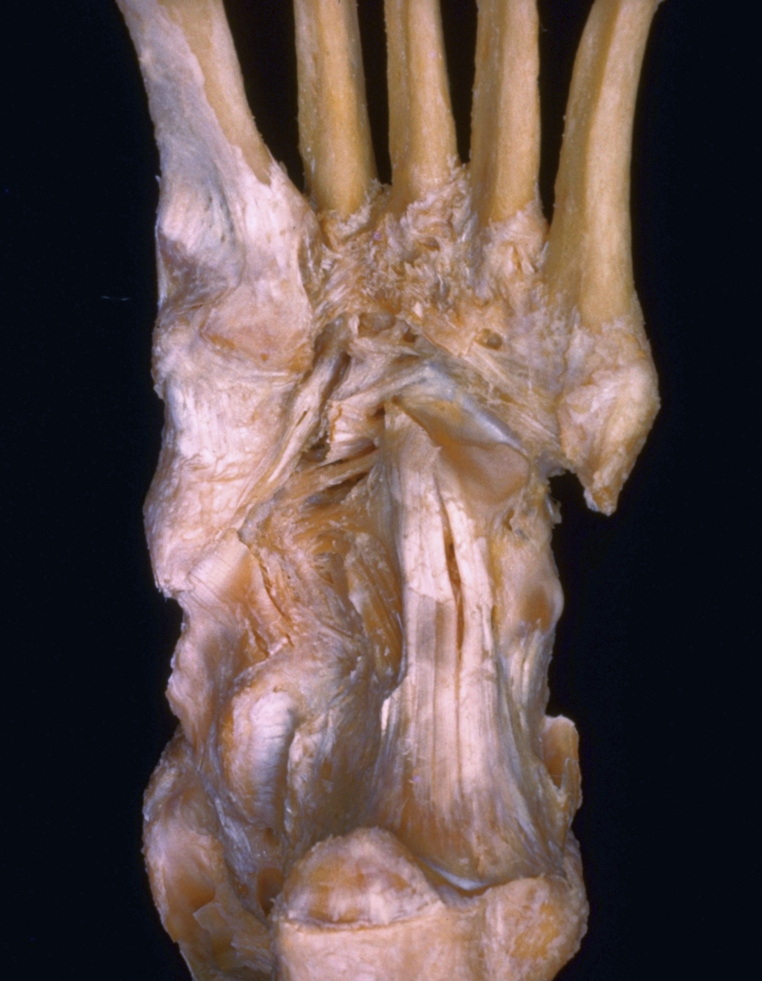
Joints of left ankle and foot
Ligaments of plantar aspect of foot, deep layer
The tendons of the peroneus longus and tibialis posterior muscles have been cut off at their insertions (1, 3, 7) to permit the deeper-lying ligaments to be brought to view. The groove marking the course of the peroneus longus tendon across the plantar part of the foot can be readily traced in the specimen (13). Facets on the calcaneus and cuboid bone are visible along the path occupied by this tendon.
- Peroneus longus muscle (insertion on 1st metatarsal)
- Plantar tarsometatarsal ligament
- Tibialis posterior muscle (part of insertion)
- Medial cuneiform bone
- Plantar cuneonavicular ligament
- Plantar cuboideonavicular ligament
- Tibialis posterior muscle (principal insertion on navicular bone)
- Plantar calcaneonavicular ligament (spring ligament)
- Sustentaculum tali
- Medial malleolus
- Medial process of tuberosity of calcaneus
- Plantar metatarsal ligaments
- Groove for peroneus longus tendon
- Tuberosity of cuboid bone
- Calcaneocuboid plantar ligament
- Long plantar ligament
- Inferior peroneal retinaculum