Joints of left ankle and foot
Ligaments of planar aspect of foot
Stanford holds the copyright to the David L. Bassett anatomical images and has assigned
Creative Commons license Attribution-Share
Alike 4.0 International to all of the images.
For additional information regarding use and permissions,
please contact the Medical History Center.
Image #202-5


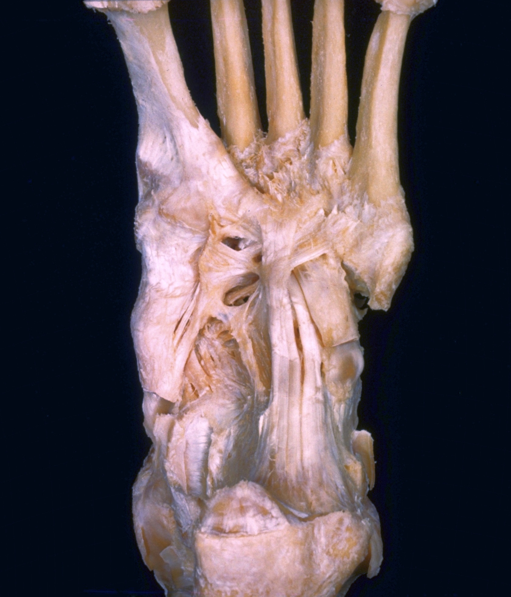
Joints of left ankle and foot
Ligaments of planar aspect of foot
In the dissection of the left foot shown in this photograph and in the subsequent views of this series the muscles, nerves and blood vessels have been entirely removed to illustrate the ligaments and joints of the ankle and foot. In connection with the study of these structures reference should also be made to view 198-4 which illustrates the ligaments on the dorsum of the foot and to view 202-4 in which the metatarsophalangeal joint of the great toe has been opened.
- Interosseous metatarsal space
- Metatarsal bone
- Area of origin of interosseous muscle
- Tendon of peroneus longus muscle
- Medial cuneiform bone (covered by ligaments)
- Plantar cuboideonavicular ligament
- Tuberosity of navicular bone
- Tendon of tibialis posterior muscle
- Plantar calcaneonavicular ligament
- Sustentaculum tali (pointer indicates groove for tendon of flexor hallucis longus muscle)
- Medial malleolus (pointer indicates groove for tendon of tibialis posterior muscle)
- Upper pointer: Medial process of tuberosity of calcaneus Lower pointer: Plantar aponeurosis
- 5th metatarsal bone
- Tuberosity of 5th metatarsal bone
- Abductor digiti minimi muscle (part of insertion)
- Calcaneocuboid plantar ligament (short plantar ligament)
- Cuboid bone (surface for tendon of peroneus longus)
- Long plantar ligament
- Inferior peroneal retinaculum
- Lateral tubercle process of calcaneus bone
- Superior peroneal retinaculum (pointer also indicates lateral malleolus)