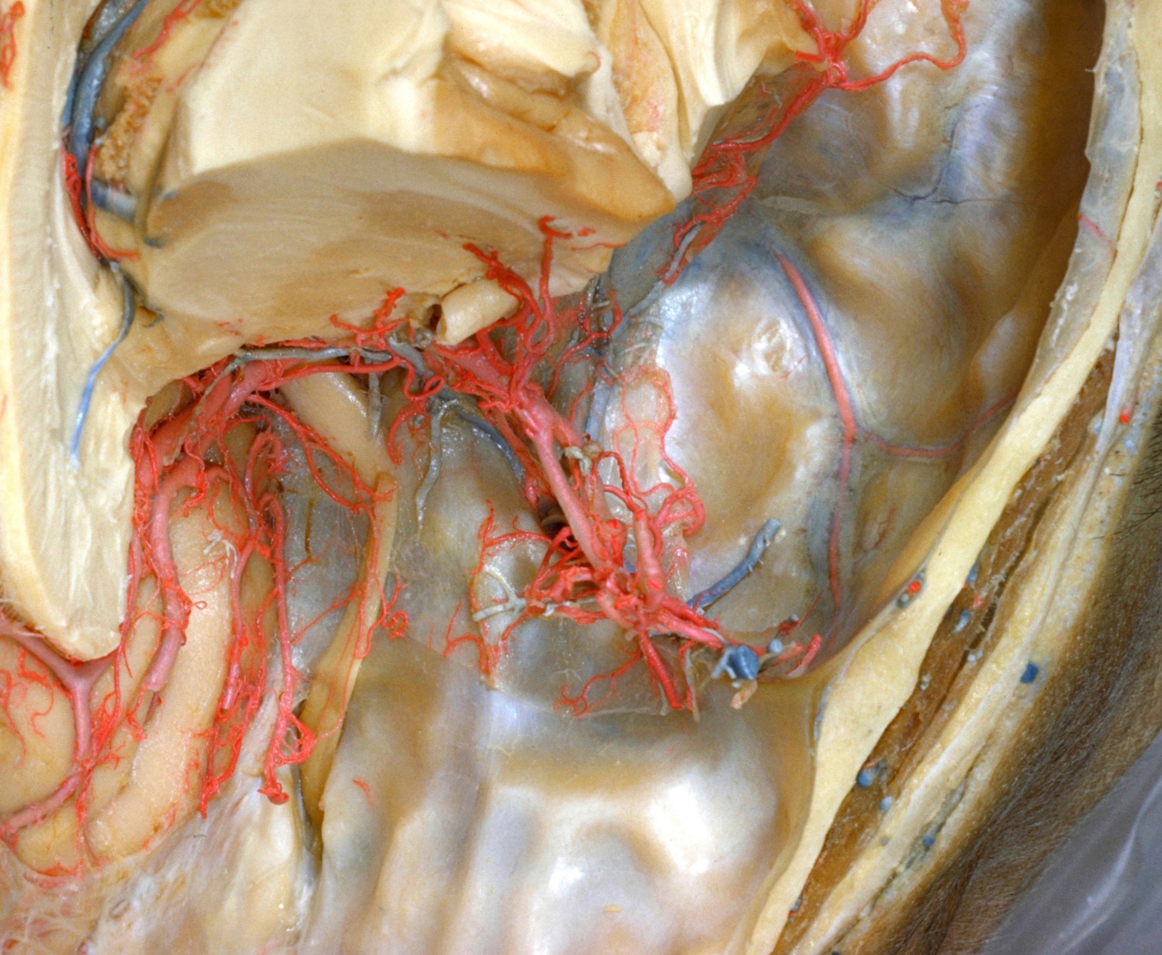
Exploration of the meninges and brain in situ
Striate arteries, anterior commissure and optic nerve viewed from above and in front
Stanford holds the copyright to the David L. Bassett anatomical images and has assigned
Creative Commons license Attribution-Share
Alike 4.0 International to all of the images.
For additional information regarding use and permissions,
please contact the Medical History Center.
Image #2-6
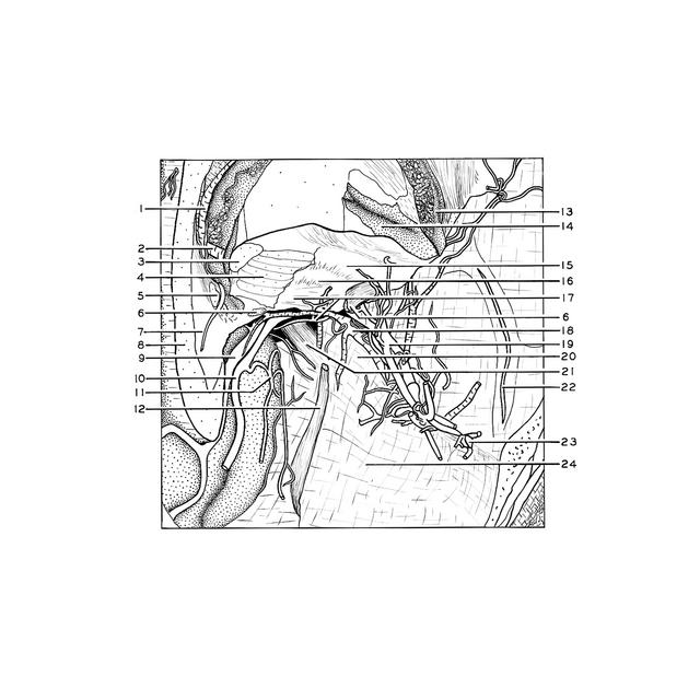
Exploration of the meninges and brain in situ
Striate arteries, anterior commissure and optic nerve viewed from above and in front
More tissue has been cut away from the basal region of the left hemisphere. The anterior commissure (6) has been partly removed. The striate arteries and the recurrent branch of the anterior cerebral artery (17) are shown in their relation to the lentiform nucleus. The temporal pole has been removed from the middle cranial fossa.
- Internal cerebral vein left
- V. terminalis
- Caudate nucleus (cut across)
- Frontal part of internal capsule (cut across)
- Fornix (column) left (cut across)
- Anterior commissure
- Septum pellucidum (note: septal vein)
- Corpus callosum
- Anterior cerebral artery right
- Anterior cerebral artery left (cut across)
- Orbital branch of anterior cerebral artery
- Olfactory tract (cut across)
- Hippocampus
- Insula
- Putamen
- External division of globus pallidus
- Internal division of globus pallidus and recurrent branch of anterior cerebral artery
- Inferior cerebral vein
- Middle cerebral artery
- Lesser wing sphenoid bone (covered by dura mater and arachnoid)
- Optic nerve (II)
- Middle meningeal artery
- Middle cerebral vein
- Anterior cranial fossa