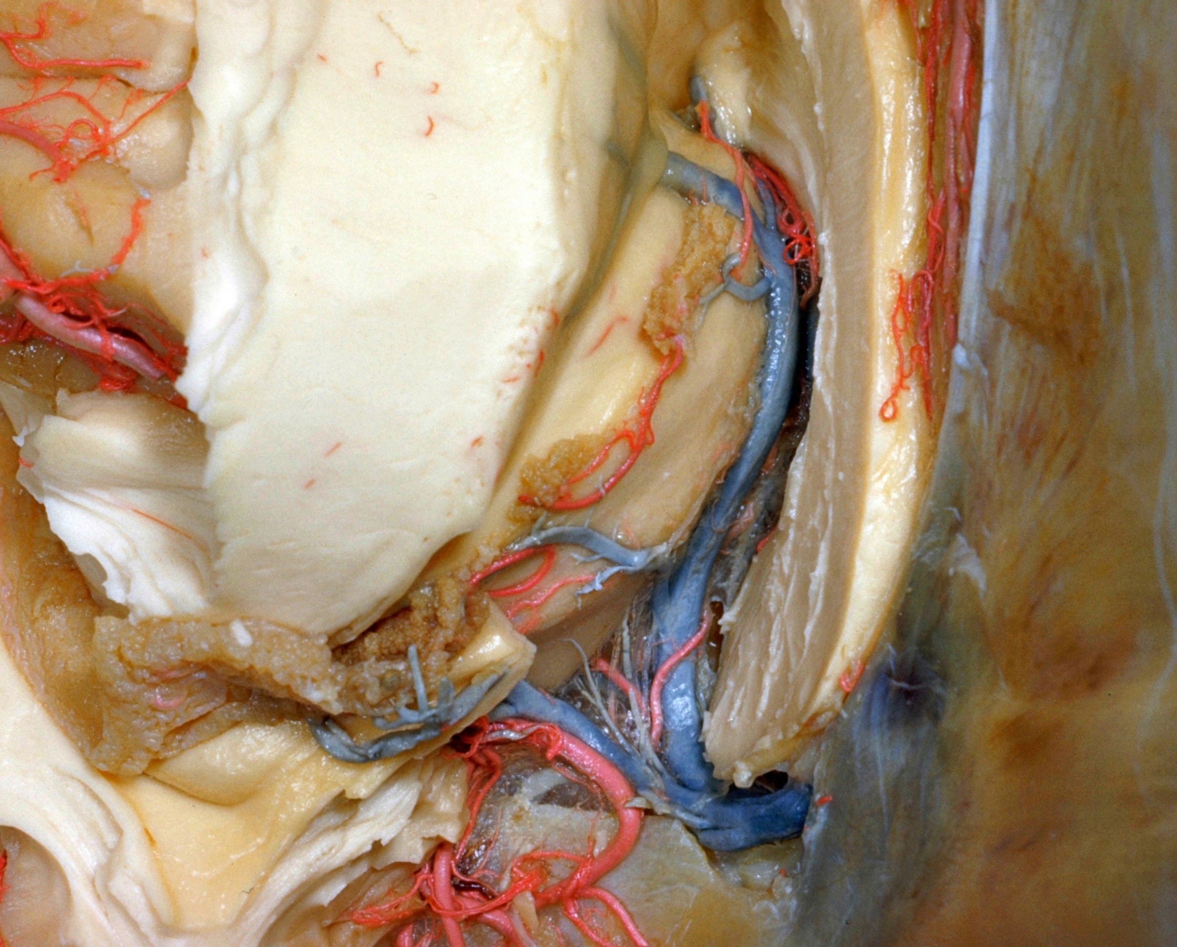
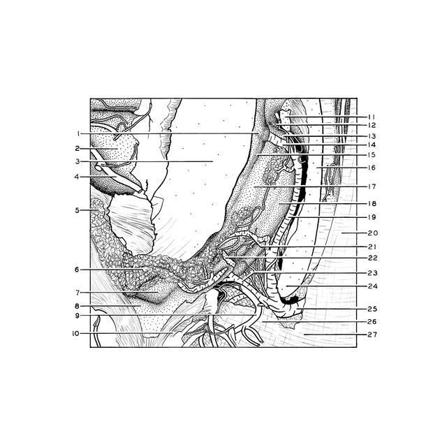
Exploration of the meninges and brain in situ
Close-up view of tributaries of internal cerebral vein
Stanford holds the copyright to the David L. Bassett anatomical images and has assigned
Creative Commons license Attribution-Share
Alike 4.0 International to all of the images.
For additional information regarding use and permissions,
please contact the Medical History Center.
Image #2-4
Exploration of the meninges and brain in situ
Close-up view of tributaries of internal cerebral vein
The left half of the fornix has been partly removed. The cut ends of the remaining parts are visible at 11 and 22. The inferior sagittal sinus lies within the falx (20) near its free margin.
- Caudate nucleus (note branches of terminal vein extending over this nucleus)
- Insula
- Emerging fibers of internal capsule (cut across)
- Posterior parietal branch of middle cerebral artery (cut off)
- Choroid plexus lying within inferior horn of lateral ventricle
- Choroidal glomus (retracted upward)
- Hippocampus
- Collateral trigone
- Branch posterior cerebral artery
- Medullary substance of occipital lobe
- Fornix (column) (cut across)
- Interventricular foramen (of Monro)
- Anterior tubercle of thalamus
- Terminal vein
- Stria terminalis (note that this passes over the vena terminalis)
- Corpus callosum (trunk) (cut through in midline)
- Lamina affixa
- Superior surface of thalamus
- Internal cerebral vein (left)
- Falx cerebri
- Choroidal branch of internal cerebral vein
- Fornix (crus) (cut across)
- Branch of internal cerebral vein from corpus callosum and roof of lateral ventricle (cut ends displaced downward)
- Corpus callosum (splenium)
- Great cerebral vein (of Galen) (seen through cut in margin of tentorium)
- Tentorium cerebelli
- Position of straight sinus within tentorium