Exploration of the meninges and brain in situ
Lateral ventricle viewed from behind; left internal cerebral vein
Stanford holds the copyright to the David L. Bassett anatomical images and has assigned
Creative Commons license Attribution-Share
Alike 4.0 International to all of the images.
For additional information regarding use and permissions,
please contact the Medical History Center.
Image #2-3
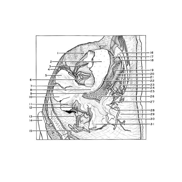
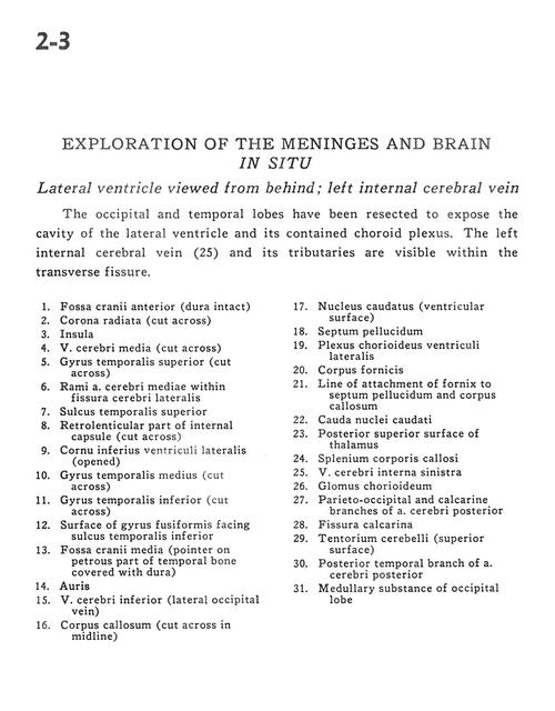
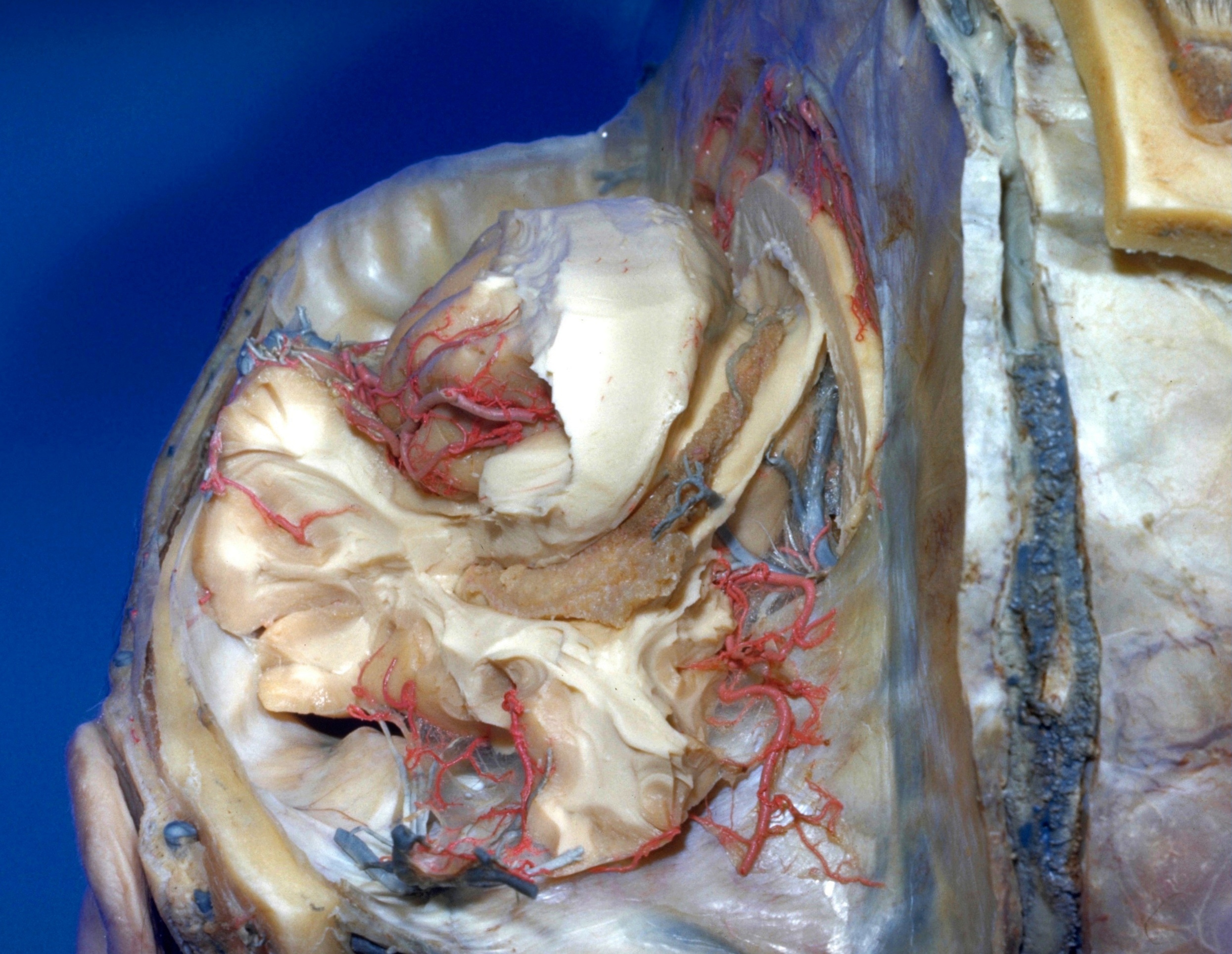
Exploration of the meninges and brain in situ
Lateral ventricle viewed from behind; left internal cerebral vein
The occipital and temporal lobes have been resected to expose the cavity of the lateral ventricle and its contained choroid plexus. The left internal cerebral vein (25) and its tributaries are visible within the transverse fissure.
- Anterior cranial fossa (dura intact)
- Corona radiata (cut across)
- Insula
- Middle cerebral vein (cut across)
- Superior temporal gyrus (cut across)
- Branch middle cerebral artery within lateral cerebral fissure
- Superior temporal sulcus
- Retrolenticular part of internal capsule (cut across)
- Inferior horn of lateral ventricle (opened)
- Medial temporal gyrus (cut across)
- Inferior temporal gyrus (cut across)
- Surface of fusiform gyrus facing inferior temporal sulcus
- Middle cranial fossa (pointer on petrous part of temporal bone covered with dura)
- Auricle
- Inferior cerebral vein (lateral occipital vein)
- Corpus callosum (cut across in midline)
- Caudate nucleus (ventricular surface)
- Septum pellucidum
- Choroid plexus lateral ventricle
- Fornix (body)
- Line of attachment of fornix to septum pellucidum and corpus callosum
- Caudate nucleus (tail)
- Posterior superior surface of thalamus
- Corpus callosum (splenium)
- Internal cerebral vein left
- Choroidal glomus
- Parieto-occipital and calcarine branches of posterior cerebral artery
- Calcarine fissure
- Tentorium cerebelli (superior surface)
- Posterior temporal branch of posterior cerebral artery
- Medullary substance of occipital lobe