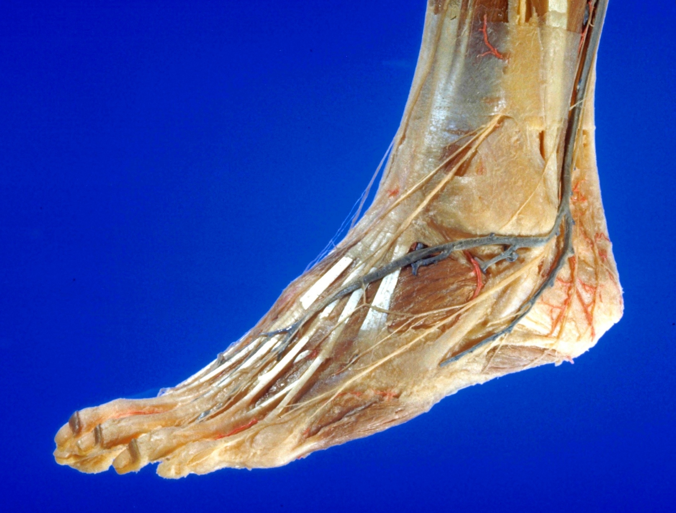
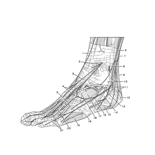
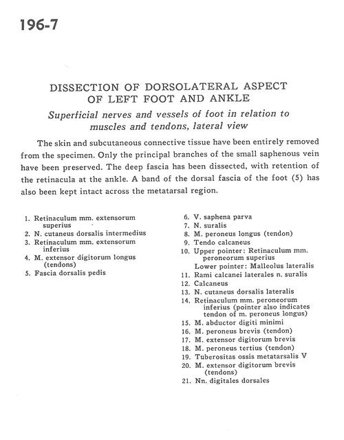
Dissection of dorsolateral aspect of left foot and ankle
Superficial nerves and vessels of foot in relation to muscles and tendons, lateral view
Stanford holds the copyright to the David L. Bassett anatomical images and has assigned
Creative Commons license Attribution-Share
Alike 4.0 International to all of the images.
For additional information regarding use and permissions,
please contact the Medical History Center.
Image #196-7
Dissection of dorsolateral aspect of left foot and ankle
Superficial nerves and vessels of foot in relation to muscles and tendons, lateral view
The skin and subcutaneous connective tissue have been entirely removed from the specimen. Only the principal branches of the small saphenous vein have been preserved. The deep fascia has been dissected, with retention of the retinacula at the ankle. A band of the dorsal fascia of the foot (5) has also been kept intact across the metatarsal region.
- Superior extensor retinaculum
- Dorsal intermediate cutaneous nerve
- Inferior extensor retinaculum
- Extensor digitorum longus muscle (tendons)
- Fascia of dorsalis pedis
- Lesser saphenous vein
- Sural nerve
- Peroneus longus muscle (tendon)
- Tendo calcaneus
- Upper pointer: Superior peroneal retinaculum Lower pointer: Lateral malleolus
- Lateral calcaneal branch of sural nerve
- Calcaneus
- Dorsal lateral cutaneous nerve
- Inferior peroneal retinaculum (pointer also indicates tendon of peroneus longus muscle)
- Abductor digiti minimi muscle
- Peroneus brevis muscle (tendon)
- Extensor digitorum brevis muscle
- Peroneus tertius muscle (tendon)
- Tuberosity of 5th metatarsal bone
- Extensor digitorum brevis muscle (tendons)
- Dorsal digital nerves