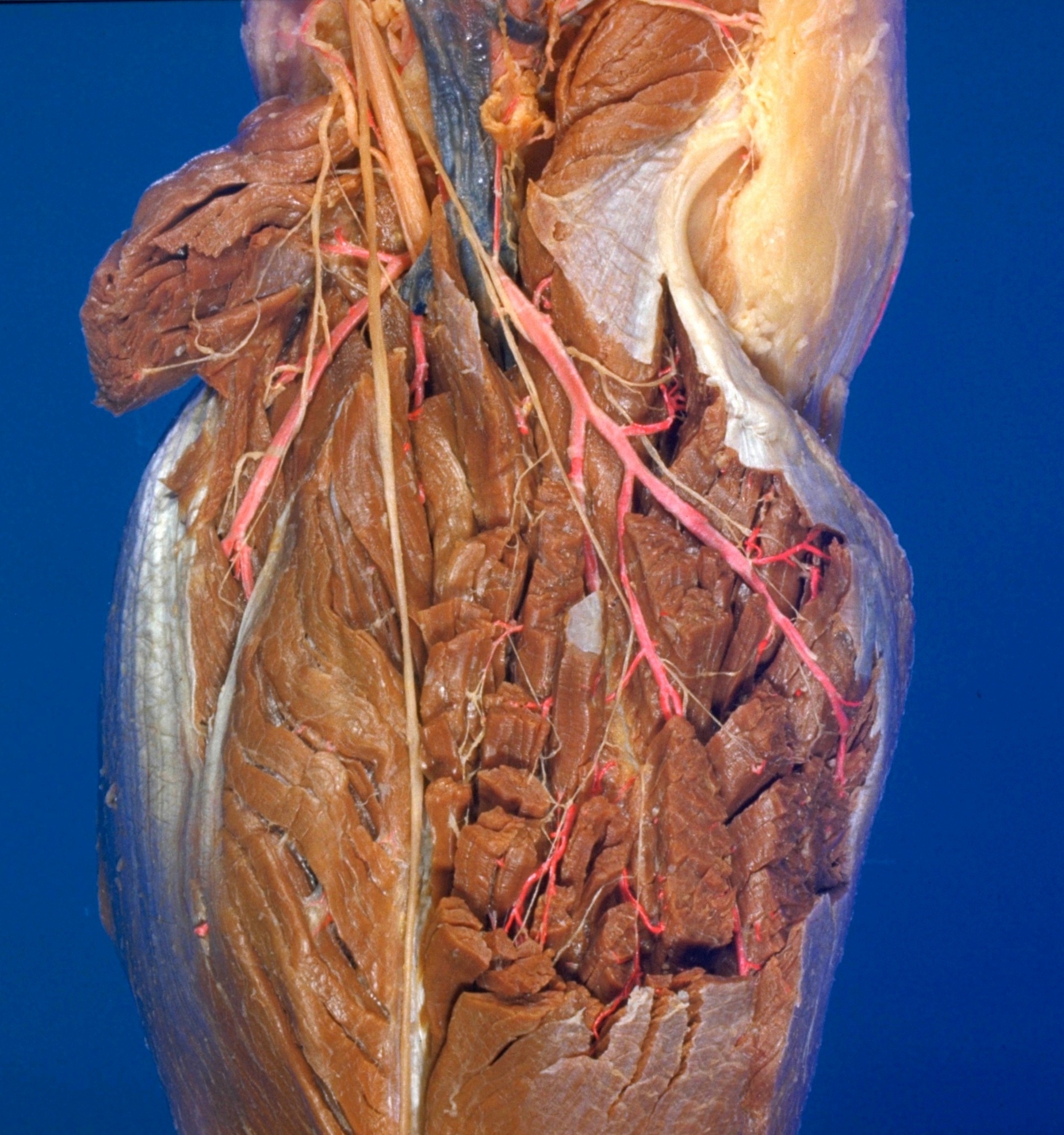
Dissection of posterior aspect of left leg
Nerve supply to gastrcnemius muscle
Stanford holds the copyright to the David L. Bassett anatomical images and has assigned
Creative Commons license Attribution-Share
Alike 4.0 International to all of the images.
For additional information regarding use and permissions,
please contact the Medical History Center.
Image #194-7
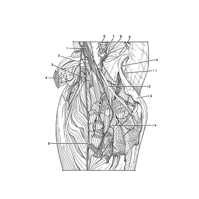
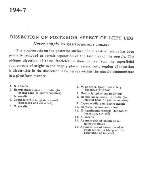
Dissection of posterior aspect of left leg
Nerve supply to gastrcnemius muscle
The aponeurosis on the posterior surface of the gastrocnemius has been partially removed to permit separation of the fascicles of the muscle. The oblique direction of these fascicles in their course from the superficial aponeurosis of origin to the deeply placed aponeurotic tendon of insertion is discernible in the dissection. The nerves within the muscles communicate in a plexiform manner.
- Tibial nerve
- Muscular branch of tibial nerve (to lateral head of gastrocnemius)
- Sural artery
- Lateral head of gastrocnemius muscle (dissected and elevated)
- Sural nerve
- Popliteal vein (popliteal artery obscured by vein)
- Popliteal lymph node
- Muscular branch of tibial nerve (to medial head Of gastrocnemius)
- Medial head of gastrocnemius muscle
- Bursa of semimembranosus muscle
- Semimembranosus muscle (tendon of insertion, cut off)
- Sural artery
- Aponeurosis of origin of gastrocnemius muscle
- Aponeurosis of insertion of gastrocnemius muscle (deep within substance of muscle)