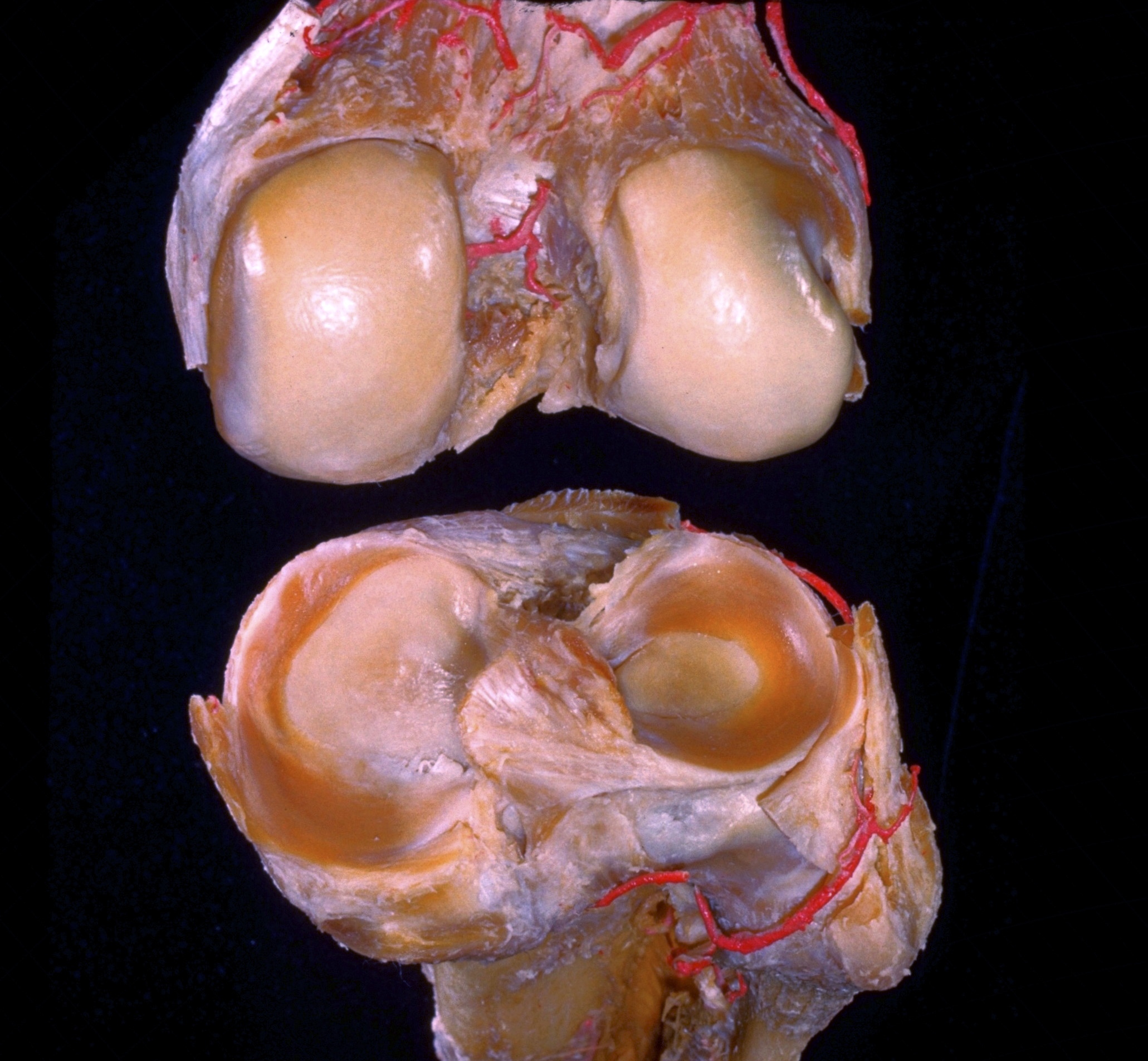
Dissection of knee
Interior of right knee joint, posterosuperior view with bones separated
Stanford holds the copyright to the David L. Bassett anatomical images and has assigned
Creative Commons license Attribution-Share
Alike 4.0 International to all of the images.
For additional information regarding use and permissions,
please contact the Medical History Center.
Image #191-5
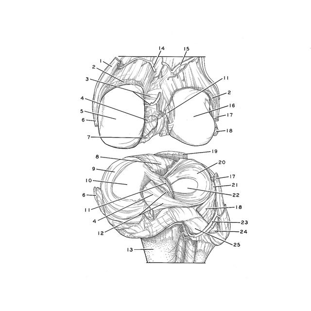
Dissection of knee
Interior of right knee joint, posterosuperior view with bones separated
The femur and tibia have been completely disarticulated. The tibia is viewed from above and behind. The femur has been pulled forward to expose its posteroinferior aspect.
- Adductor magnus muscle (tendon cut at insertion)
- Capsule of knee joint
- Upper pointer: Intercondylar line Lower pointer: Medial genicular artery
- Posterior cruciate ligament (divided)
- Medial condyle of femur
- Tibial collateral ligament
- Synovial membrane
- Anterior intercondylar area (note the absence of a transverse ligament in this specimen)
- Medial meniscus
- Medial condyle of tibia (articular surface)
- Anterior cruciate ligament (divided)
- Upper pointer: Anterior meniscofemoral ligament Lower pointer: Posterior meniscofemoral ligament
- Body of tibia
- Medial superior genicular artery
- Lateral superior genicular artery
- Lateral condyle of femur
- Collateral ligament of fibula (divided)
- Popliteus muscle (tendon of origin)
- Patellar ligament (cut off)
- Lateral meniscus
- Articular surface on inner surface of tendon of popliteus muscle
- Lateral condyle of tibia (articular surface)
- Lateral inferior genicular artery
- Popliteal arcuate ligament (cut at attachment to apex of fibula)
- Subpopliteal recess