Dissection of anterior and medial aspects of thigh
Nerve supply to adductor brevis muscle; medial femoral circumflex artery
Stanford holds the copyright to the David L. Bassett anatomical images and has assigned
Creative Commons license Attribution-Share
Alike 4.0 International to all of the images.
For additional information regarding use and permissions,
please contact the Medical History Center.
Image #188-2
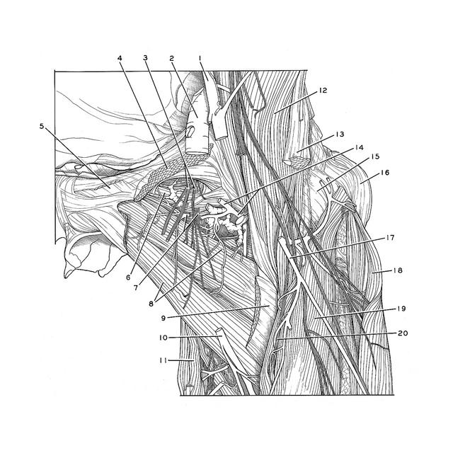
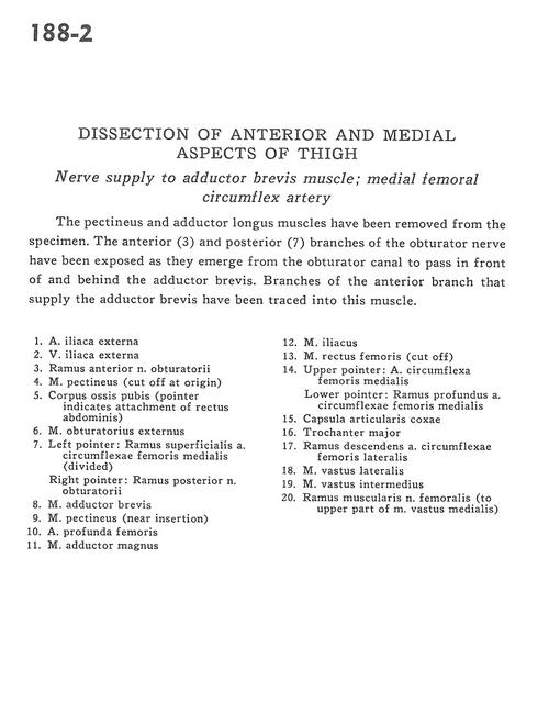
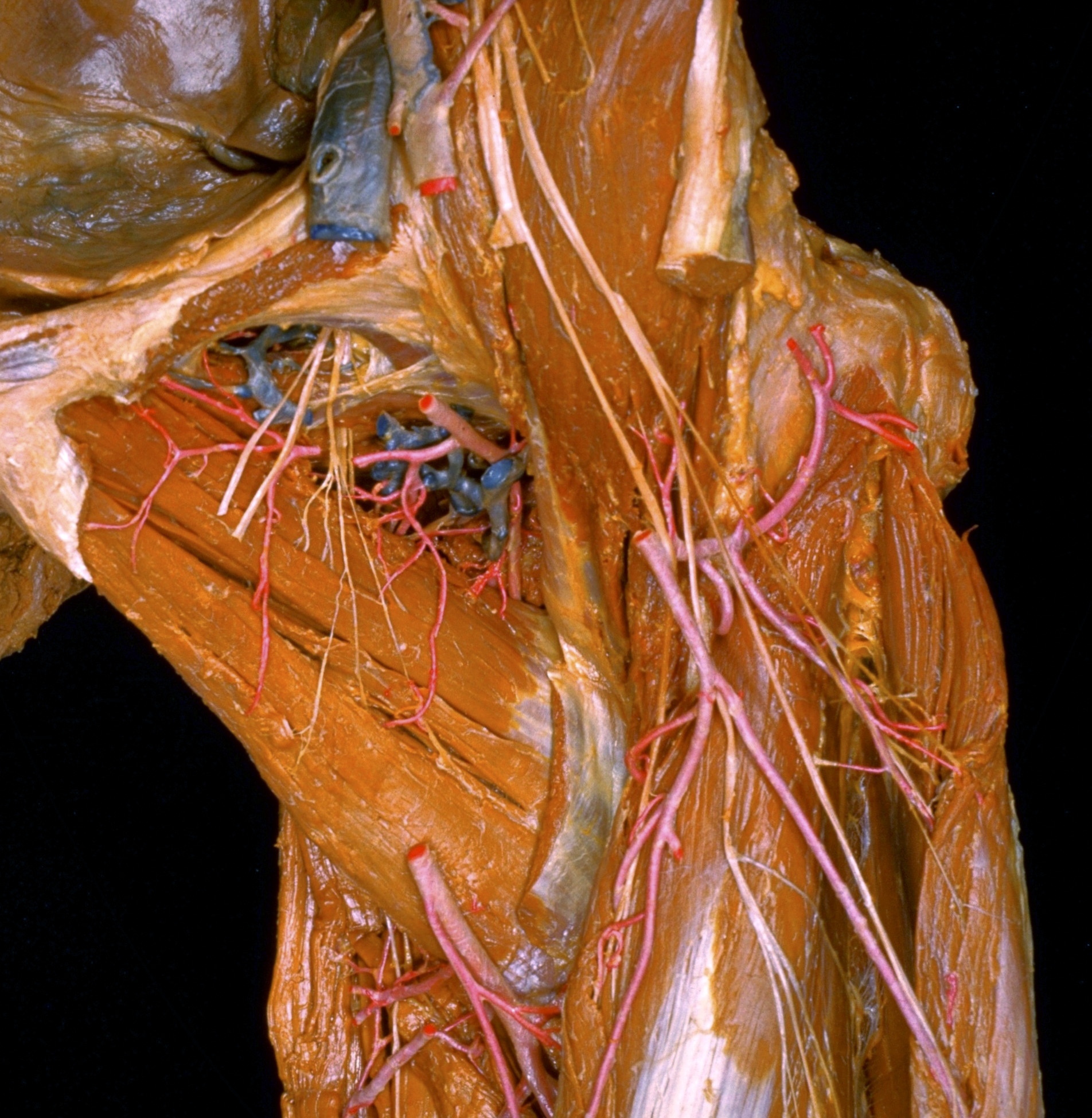
Dissection of anterior and medial aspects of thigh
Nerve supply to adductor brevis muscle; medial femoral circumflex artery
The pectineus and adductor longus muscles have been removed from the specimen. The anterior (3) and posterior (7) branches of the obturator nerve have been exposed as they emerge from the obturator canal to pass in front of and behind the adductor brevis. Branches of the anterior branch that supply the adductor brevis have been traced into this muscle.
- External iliac artery
- External iliac vein
- Anterior branch of obturator nerve
- Pectineus muscle (cut off at origin)
- Body of pubis (pointer indicates attachment of rectus abdominis)
- Obturator externus muscle
- Left pointer: Superficial branch of medial circumflex artery (divided) Right pointer: Posterior branch of obturator nerve
- Adductor brevis muscle
- Pectineus muscle (near insertion)
- Deep femoral artery
- Adductor magnus muscle
- Iliacus muscle
- Rectus femoris muscle (cut off)
- Upper pointer: Medial femoral circumflex artery Lower pointer: Deep branch of medial femoral circumflex artery
- Hip articular capsule
- Greater trochanter
- Descending branch of lateral femoral circumflex artery
- Vastus lateralis muscle
- Vastus intermedius muscle
- Muscular branch of femoral nerve (to upper part of vastus medialis muscle)