Exploration of gluteal region and hip
Interior of left hip joint, lateral view
Stanford holds the copyright to the David L. Bassett anatomical images and has assigned
Creative Commons license Attribution-Share
Alike 4.0 International to all of the images.
For additional information regarding use and permissions,
please contact the Medical History Center.
Image #182-2
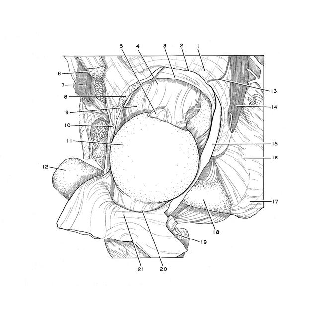
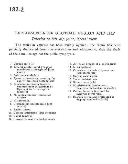
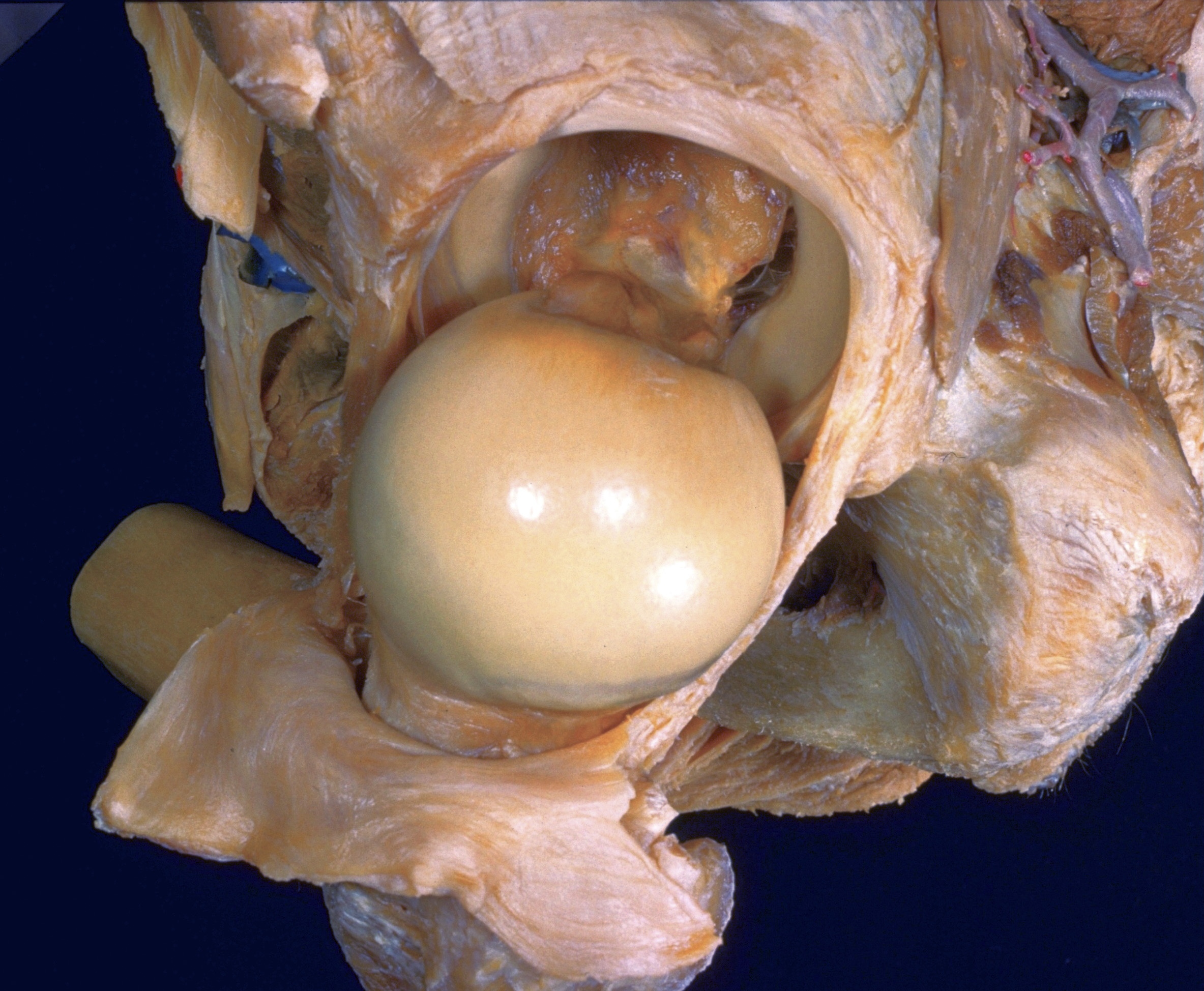
Exploration of gluteal region and hip
Interior of left hip joint, lateral view
The articular capsule has been widely opened. The femur has been partially dislocated from the acetabulum and adducted so that the shaft of the bone lies against the pubic symphysis.
- Body of ilium
- Line of reflection of synovial membrane at margin of joint cavity
- Acetabular margin
- Synovial membrane covering fat pad within acetabular fossa
- Ligament of femoral head (pointer near attachment of ligament to fovea of head of femur)
- Rectus femoris muscle (tendon of origin)
- Femoral nerve
- Iliofemoral ligament (cut across)
- Acetabular lunate surface
- Articular capsule (cut through)
- Head of femur
- Body of femur (in background)
- Articular branch of sciatic nerve
- Sciatic nerve
- Articular capsule (ischiofemoral ligament)
- Body of ischium
- Ischial tuberosity
- Ischial ramus
- Piriform muscle (tendon near insertion on greater trochanter)
- Neck of femur (covered by synovial membrane)
- Articular capsule (reflected to display zona orbicularis)